Dear MRI Insider,
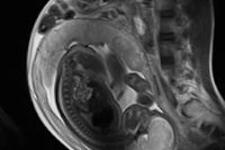
Fetal MRI is gaining clinical acceptance and being used more extensively across Europe, but there's still an urgent need to optimize and standardize patient care and imaging parameters to realize its potential, avoid unnecessary repeat examinations, and improve image quality and efficiency, according to a team of researchers from a top London facility.
Earlier this week, the group spelt out exactly how they have achieved success in fetal MRI. Make sure you don't miss their practical advice. To read more, click here.
Bone metastases cause severe pain and rapid degradation in quality of life in patients with advanced cancer. Such metastases are usually treated using radiation therapy, but this isn't suitable, or successful, for all patients. Another option may be MR-guided focused ultrasound. Click here to learn more.
Breast MRI was high on the agenda at the 95th congress of the German Radiology Society, the DRK, organized jointly this year with the Austrian Radiology Society. The highly respected Dr. Christiane Kuhl from Aachen was one of the expert speakers. To find out more, click here.
Also, we've posted news reports from the DRK about the use of MRI in diagnosing nondescript chest pain and detecting endometriomas.
Increasing numbers of extremely old and frail patients are being referred for MRI examinations, but is it always appropriate for them to undergo such procedures? Dr. Paul McCoubrie considers this question in his latest column. Get the story here.
This letter features only a small selection of the many articles posted in the MRI Digital Community. Check out the rest of the full list below this message.


![Overview of the study design. (A) The fully automated deep learning framework was developed to estimate body composition (BC) (defined as subcutaneous adipose tissue [SAT] in liters; visceral adipose tissue [VAT] in liters; skeletal muscle [SM] in liters; SM fat fraction [SMFF] as a percentage; and intramuscular adipose tissue [IMAT] in deciliters) from MRI. The fully automated framework comprised one model (model 1) to quantify different BC measures (SAT, VAT, SM, SMFF, and IMAT) as three-dimensional (3D) measures from whole-body MRI scans. The second model (model 2) was trained to identify standardized anatomic landmarks along the craniocaudal body axis (z coordinate field), which allowed for subdividing the whole-body measures into different subregions typically examined on clinical routine MRI scans (chest, abdomen, and pelvis). (B) BC was quantified from whole-body MRI in over 66,000 individuals from two large population-based cohort studies, the UK Biobank (UKB) (36,317 individuals) and the German National Cohort (NAKO) (30,291 individuals). Bar graphs show age distribution by sex and cohort. BMI = body mass index. (C) After the performance assessment of the fully automated framework, the change in BC measures, distributions, and profiles across age decades were investigated. Age-, sex-, and height-adjusted body composition reference curves were calculated and made publicly available in a web-based z-score calculator (https://circ-ml.github.io).](https://img.auntminnieeurope.com/mindful/smg/workspaces/default/uploads/2026/05/body-comp.XgAjTfPj1W.jpg?auto=format%2Ccompress&fit=crop&h=100&q=70&w=100)


![Overview of the study design. (A) The fully automated deep learning framework was developed to estimate body composition (BC) (defined as subcutaneous adipose tissue [SAT] in liters; visceral adipose tissue [VAT] in liters; skeletal muscle [SM] in liters; SM fat fraction [SMFF] as a percentage; and intramuscular adipose tissue [IMAT] in deciliters) from MRI. The fully automated framework comprised one model (model 1) to quantify different BC measures (SAT, VAT, SM, SMFF, and IMAT) as three-dimensional (3D) measures from whole-body MRI scans. The second model (model 2) was trained to identify standardized anatomic landmarks along the craniocaudal body axis (z coordinate field), which allowed for subdividing the whole-body measures into different subregions typically examined on clinical routine MRI scans (chest, abdomen, and pelvis). (B) BC was quantified from whole-body MRI in over 66,000 individuals from two large population-based cohort studies, the UK Biobank (UKB) (36,317 individuals) and the German National Cohort (NAKO) (30,291 individuals). Bar graphs show age distribution by sex and cohort. BMI = body mass index. (C) After the performance assessment of the fully automated framework, the change in BC measures, distributions, and profiles across age decades were investigated. Age-, sex-, and height-adjusted body composition reference curves were calculated and made publicly available in a web-based z-score calculator (https://circ-ml.github.io).](https://img.auntminnieeurope.com/mindful/smg/workspaces/default/uploads/2026/05/body-comp.XgAjTfPj1W.jpg?auto=format%2Ccompress&fit=crop&h=112&q=70&w=112)


