Interventional radiology was truly the focus of the 95th German Congress of Radiology (DRK), and as the second part of this year´s motto warned us, "Radiology is (...) treatment." To help protect the environment and due to the omnipresent Internet, the organizers decided not to distribute a print version of the book of abstracts. They rather provided free WLAN at the Congress Center Hamburg with access to the online program planner. Yet no matter how you studied the program, the immense role of interventional radiology within our specialty was obvious everywhere.
Newcomers and expert interventional radiologists could choose between basic lectures given by respected authorities or more interactive seminars. Numerous scientific sessions were organized, presenting the newest developments within interventional radiology, and the latest devices could be inspected at the large exhibition area. Last but not least, workshops were offered to train delegates in endovascular procedures. These were organized by the German Society of Interventional Radiology (Deutsche Gesellschaft für Interventionelle Radiologie, DeGIR) and provided structured teaching in different modules, such as endovascular recanalization and embolization as well as tumor ablation. After successful completion, participants could acquire several certificates documenting their area and level of experience.
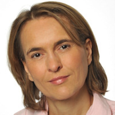
 Hamburg will again host the DRK in 2015, but Leipzig will be the venue for the congress from 2016. Image courtesy of Michael Reiter.
Yet, we should not forget the first part of this year´s motto: "Radiology is diagnosis." Noteworthy, one special focus was set by the organizers on x-ray. Every day numerous sessions were designed to refresh our knowledge of this classic, but sometimes underestimated and even neglected, imaging technique. Particularly in the young generation of radiologists there is a high risk that the immense diagnostic information provided by plain radiography gets lost, according to the organizers. Not surprisingly, numerous lectures were addressed to junior radiologists discussing virtually all aspects of plain radiography, from the physical basis to legal issues of radiation protection, and finally covering every possible clinical application.
The next generation
As in previous years, the organizers continued to invest in youth and invited highly motivated medical students to the meeting. The initiative known as "die hellsten Köpfe für die Radiologie" ("Brightest minds for radiology") provided travel grants for selected students. For these younger delegates, a special infrastructure was designed, including the students' lounge on the 1st floor, providing an area to relax and to network.
Similar to the RSNA and ECR, snacks and drinks were offered to get ready for special focus sessions and for the Sono4you project. The latter was designed as an interactive workshop providing basic skills in ultrasound within a multisession course. During the two "young investigator award" sessions, the most promising scientific research was presented. The judges had a really tough job to decide which of the excellent candidates would get the much sought after award.
Martin Krämer from the medical physics group at Jena University Hospital (chair: Dr. Jürgen Reichenbach) and Dr. Julian Luetkens from the clinic of radiology at the Bonn University Hospital (chair: Dr. Hans Schild) were chosen. Krämer received an award for his paper titled "Intrinsically gated cardiac MR imaging of the rat at a clinical 3T scanner." His approach requires neither special software nor a dedicated animal scanner, but still provides high temporal and spatial resolution. Luetkens presented his clinical work, "Diagnostic performance of multiparametric cardial MRI (...) in acute myocarditis at 3 Tesla." He demonstrated the significant additional value of advanced MRI methods for differential diagnosis of myocardial disease, and reported that most of all T1 relation times proved to be most helpful to diagnosis of acute myocarditis.
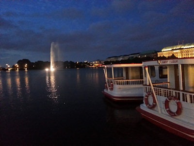 Nightlife in Hamburg: After a tough congress day, there are plenty of places to relax in the maritime capital of the north. One option is very close to the congress venue: The "Binnenalster" ("Inner Alster Lake"), an artificial lake within the city center. Image courtesy of Dr. Matthias Dietzel.
Breast MRI gathers momentum
Independent from the annually changing motto, breast imaging has traditionally been a central theme of the DRK. In Hamburg, the most prominent breast radiologists from Austria and Germany provided state-of-the-art teaching sessions and shared their visions on up-to-date patient care.
One highlight was certainly the workshop on clinical breast MRI moderated by Dr. Michael Fuchsjäger from Graz, Austria. Empty seats during the closing sessions of any congress are a well-known phenomenon. Who would blame delegates, exhaustedly travelling home while the final sessions are still running? Nevertheless, this session was sold out, and delegates were even standing to join this important workshop. Certainly it was both the prominent speakers and the topic that attracted delegates to join this furious finale of the congress.
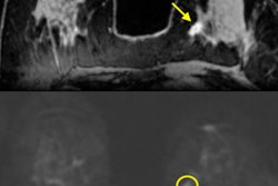
Giving an excellent overview on the potential of breast MRI to assess in situ cancers, Dr. Wolfgang Buchberger from Innsbruck started the session. Traditionally there have been many myths on the assessment of ductal carcinoma in situ (DCIS) using MRI. However, since the original Lancet paper of Dr. Christiane Kuhl from Bonn (now based in Aachen) was published in 2007, there are clear data proving that breast MRI has an excellent sensitivity to detect this disease. A notable strength of this method is the assessment of biologically aggressive, high-grade DCIS lesions. In this clinically most relevant subgroup, MRI even shows a higher sensitivity compared with conventional mammography, Buchberger concluded.
"One of the biggest troublemakers in breast MRI are nonmass enhancements (NMEs)" stated Dr. Pascal Baltzer from the University of Vienna in his precise and comprehensive presentation. "These are frequent types of lesions and BI-RADS criteria are of little use for the differential diagnosis between malignant and benign NMEs." Interestingly, this fact is still rarely recognized within the breast MR community and advanced techniques, e.g. diffusion-weighted imaging and spectroscopy, contribute little to this diagnostic dilemma; this is probably most likely due to their intrinsically lower spatial resolution and to partial volume effects which are almost always present in NME due to diffuse growth patterns, he added.
 Lining up for the big event: Delegates wait patiently in line for a very popular session. Image courtesy of Dr. Matthias Dietzel.
One possible approach to address this challenge is the use of additional diagnostic features. Citing his own group's publications and giving clear examples, Baltzer recommended including high resolution nonfat-saturated T2-weighted sequences into every clinical MRI scanning protocol. Such images provide additional details on NMEs, e.g., perifocal edema, cystic components, ductal obstruction, and fibrotic foci, all of which could significantly enhance diagnostic performance, according to Baltzer, who summarized the current status of his research and clinical experience.
Preoperative staging remains one of the major indications for breast MRI. Kuhl gave an enthusiastic and provocative lecture on this topic, sharing her own views on the current controversial debate. First of all, there is convincing evidence that breast MRI identifies a significantly higher tumor burden compared with conventional assessments. This is not questioned, even by the critics of this imaging method: compared with the conventional work-up, breast MRI shows a more accurate assessment of the tumor size and identifies additional tumor foci in the ipsilateral (multifocal or multicentric disease) or contralateral breast in a significant proportion of the patients.
Nevertheless, current German S3 guidelines do not recommend routine use of breast MRI in the preoperative setting. Critics argue that there are no prospectively randomized controlled trials proving a positive effect on patient outcome, if breast MRI is compared directly with conventional assessment. Such data would be desirable, no question. "Yet, it would take decades to get clear evidence on this. We cannot wait until we have these data," Kuhl said. "Our gynecologist and oncologist would not randomize their patients, because the benefit of preoperative setting is absolutely clear to them."
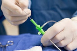
Yet, until your clinical partners are convinced by the method, three basic prerequisites have to be met. Firstly, the imaging site needs specially trained MR radiologists, and a high case load is a must, otherwise you won't do a good job, which is similar to mammography. Secondly, with lesions detected using MRI only, you still need MR-guided procedures readily available on your site because otherwise clear histological sampling won't be possible. Thirdly, for the interdisciplinary side, there is a need for clear communication of radiological findings to the surgeon, and this is often underestimated.
Without regular face-to-face case discussions, it is difficult to translate such complicated MRI findings into personalized therapy planning. According to Kuhl, all these criteria were not met in the only major randomized controlled trial on this subject, the COMICE trial.
Finally, Kuhl discussed whether prospective randomized controlled trials are really necessary for preoperative staging. Citing the Oxford evidence-based medicine criteria, this would not ultimately be necessary per se, she stated. And -- most interestingly -- "There is no single study with a similar rational investigating breast ultrasound or mammography. And no one requires it," she said.
Being also a board-certified neuroradiologist, Kuhl cited the neuro-oncology literature. "There are no prospective randomized controlled trials on preoperative MRI of glioblastoma, proving the benefit on patient outcome compared to other/cheaper imaging methods (e.g. CT). Moreover, everybody does it and no one questions this approach because the benefit is clear."
Dr. Thomas Helbich from the University of Vienna then gave a thorough overview on the assessment of therapy response in patients receiving neoadjuvant chemotherapy. Of course, one could simply measure the change of tumor dimensions during course of treatment, but it is probably here where advanced MRI techniques, including pharmacokinetic modelling, diffusion and spectroscopy, will be most beneficial for the patient. Such methods help the radiologist to perform functional assessment of the tumor and provide a more personalized approach to patient treatment. "If you go home, on Monday you should provide such methods to your patients," he concluded.
After four days packed with new scientific results, useful knowledge and networking, there is no doubt that the DRK was a great success. Make sure you don´t miss it next year because it will be the last meeting in the maritime capital of the north, Hamburg. Starting from 2016, Leipzig will be the host to the world's oldest meeting dedicated to medical imaging.
