A case series of 12 male retired professional motorsport drivers indicated a high prevalence of mild neurological abnormalities, with eye movement dysfunction emerging as the clearest marker of cumulative head impact exposure, researchers have reported.
Two findings were "particularly noteworthy," wrote a team led by Kévin Polet, PhD, of Princess Grace Hospital in Monaco. The group's research was published May 28 in Sports Medicine Open.
"[We found that] eye movement abnormalities correlated with cumulative motorsport exposure duration, and [that] participants showed an atypical cognitive profile with above-average visuospatial and processing speed performances," it noted. "This unique profile may make standard clinical assessments less sensitive for detecting subtle pathological processes -- whether during [racing drivers'] active career or later in life."
Professional motorsport drivers are at high risk of concussions and repeated subconcussive head impacts, Polet and colleagues explained. Concussion symptoms can persist, and repeated head impacts are thought to contribute to a "gradual accumulation of axonal damage over time," they noted. But long-term neurological consequences of practicing professional motorsports remain unclear.
The investigators analyzed a case series to determine the neurological health status of former professional motorsports drivers and to explore any relationships between cumulative head impacts exposure and long-term brain health. The research included 12 former professional motorsport drivers (all male, median age 56); of these, 58% presented with cognitive complaints and 50% with motorsport-related concerns. Mean motorsport practice duration was 31 years, while mean professional career span was 26 years and mean retirement age was 40 years. All participants had participated in multiple motorsport disciplines, and the majority (83%) had competed in open-wheel single-seater racing ("formula") and/or sport car racing (also enclosed-wheel, but two-seater vehicles).
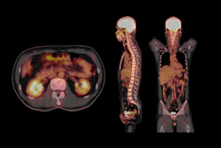
Study participants underwent both brain MRI (with SWIp sequences and diffusion tensor imaging [DTI]) and F-18 FDG-PET scans. The researchers also assessed participants' brain health through clinical assessments, including the Sport Concussion Assessment Tool 5th Edition (SCAT5) symptom inventory, neuropsychological assessment, and video-oculography.
The group found the following:
- 75% of study participants had at least one symptom on SCAT5;
- 33% had at least one cognitive impairment;
- 75% had at least one impaired eye movement;
- 58% had at least one MRI abnormality (i.e., frontal lobe atrophy and microvascular lesions);
- 92% had at least one DTI abnormality (reduced fractional anisotropy in at least one white-matter region, with reductions concentrated in the left anterior corona radiata); and
- 33% at least one 18 FDG-PET abnormality.
Interestingly, the men's attention, processing speed, visual episodic, and working memory skills were above average, the authors wrote, which they attributed to "a high cognitive reserve built through intensive motorsport practice." Only eye movement abnormalities were correlated to total exposure duration and to professional practice duration, they noted.
 Illustrations of brain MRI abnormalities observed in a population of former professional motorsport drivers. (A) A left frontal lacune observed in axial T2-weighted 3D FLAIR sequence; (B) a left frontal microbleed observed in axial SWIp sequence in a 77-year-old participant. (C) Frontal lobes atrophy and white matter hyperintensities scored 2 on Fazekas’ scale with confluent periventricular lesions and “multi-spot pattern” for the deep white matter observed in axial T2-weighted 3D FLAIR sequence in a 68-year-old participant. (D) a micronodular vacuolating neuronal tumor incidentally discovered in axial T2-weighted 3D FLAIR in 54-year-old participant. (E) Frontal and parietal lobes atrophies in a 43-year-old participant. (F) Cerebellar atrophy in a 53-year-old participant observed in sagittal T1-weighted 3D sequence.Sports Medicine Open
"These observations highlight the need for large-scale longitudinal studies that integrate pre-season baselines with video-oculography and DTI, along with prolonged post-career follow-up, to specifically investigate the effects of repeated head impacts on this population during their career and throughout aging," the team concluded.
Access the full study here.