While the technology at the high end of the MRI spectrum becomes more complex, Prof. Joan C. Vilanova, MD, PhD, of Girona University Hospital, is looking at the bottom line. In his department, musculoskeletal (MSK) imaging, specifically the lumbar spine and knee, accounts for 80% of MRI demand.
Vilanova’s data suggests that for these high-volume cases, field strength is a matter of economics rather than medicine. Across a comparison of 0.25T, 1.5T, and 3T systems, he found that the diagnostic outcome remained identical. What changed was the cost: Low-field systems operated at roughly half the cost of 1.5T units.
 "The diagnosis does not change with field strength. Only the cost does," said Prof. Joan C. Vilanova, MD, PhD of Girona University Hospital.Courtesy of Claudia Tschabuschnig
"Reimbursement stays the same regardless of which machine performs the scan," Vilanova noted in a session at ECR 2026. Under the current European healthcare pressures, the "default" move toward high-field systems for routine MSK work is becoming increasingly difficult to justify financially.
Bringing the scanner to the patient
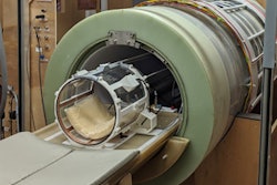
The debate over field strength isn't just about saving money; it’s about expanding the reach of the modality. Prof. Massimo Caulo, MD, of the University G. d'Annunzio of Chieti-Pescara, highlighted the clinical impact of portable, very low-field MRI.
For the most vulnerable patients -- those on ECMO, ventilators, or with pacemakers -- the journey to the radiology department is a high-risk endeavor. Transferring an unstable patient can take 90 minutes and risks tube displacement or hemodynamic instability. Portable MRI brings that time down to under 15 minutes at the bedside.
In a recent Italian trial, portable MRI demonstrated higher sensitivity than CT for detecting acute stroke in its early phases. By removing the "transportation barrier," clinicians can now access MRI for patients who were previously deemed "un-scannable."
Where 5T earns its keep
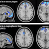
If low-field is for the routine, what is ultra-high field for? Bing Zhang, MD, PhD, of Nanjing Drum Tower Hospital, argues that the value of 5T systems is found in the "invisible" details of neuroscience.
With a 40% to 60% boost in signal-to-noise ratio over standard 3T systems, 5T imaging allows researchers to see structures like cortical layers and perivascular spaces used in glymphatic research. These systems are proving vital for detecting early cognitive decline in patients who do not yet show visible hippocampal atrophy.
However, Zhang admitted the clinical translation remains narrow. While 5T is a powerhouse for specialized neuroscience centers, its added value for routine clinical work remains marginal.
AI pros and cons
The push for faster MRI scans has made AI denoising tools a staple in modern departments. These tools work by taking a noisy, rapid acquisition and "filling in" the data to produce a clean image.
Sven Haller, MD, PhD, of the University Hospitals of Geneva, has seen this firsthand. An 18-year-old 1T open scanner, with AI acceleration applied, produced images he described as comparable to 3T. For older equipment, claustrophobic patients, and departments that cannot justify a new high-field system, the tools deliver something real. He also pointed to the ecological and economic benefits: lower energy consumption, shorter slot times, and better patient throughput on existing hardware.
 AI-based acceleration tools can make lesions disappear and even more concerning, they can invent structures that do not exist," warned Sven Haller, MD, PhD of University Hospitals of Geneva.Courtesy of Claudia Tschabuschnig
But the technology is not without risk. During his presentation, Haller shared a case: two micrometastases visible on a standard MRI scan completely disappeared on an AI-accelerated version. When the patient returned to conventional imaging, the lesions were still there.
"AI-based acceleration tools can make lesions disappear, and even more concerning, they can invent structures that do not exist," Haller said. His concern was not with the technology itself but with the absence of validation standards.
After reviewing 82 research studies on MRI acceleration, he found that not a single one tested whether the AI reconstruction had introduced findings that were not actually there, or removed findings that were. What the studies measured was image quality, signal, contrast, visual scores. Despite this, the radiologist remains legally liable if a lesion is missed.
There are no agreed criteria for how much acceleration is safe, and no standard validation process covers hallucinations.
"Hallucination is completely underdiagnosed and under-investigated," he noted.