Never offer to eat your pants – this is one of Dr. Paul McCoubrie's 100 rules of radiology. The lack of an authoritative guide or moral compass on how to become a good radiologist prompted him to compile the rules, and the welcome news is he's now completed the mammoth task.
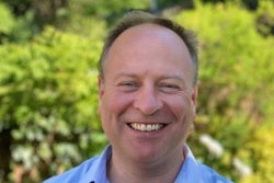
The overall aim is to provide a manifesto for radiologists across the globe to raise their game and serve patients better, says McCoubrie, a consultant radiologist at Southmead Hospital in Bristol, U.K., and regular columnist for AuntMinnieEurope.com since 2013. He looks hard at radiology but also provides "a distinctly wry look at the curious and occasionally alien world of hospital-based medical practice."
"I ask questions and poke fun, but it is serious intent," he notes. "My motto is 'first make you laugh, then make you think.' Or, as George Bernard Shaw wrote, 'My method is to take the utmost trouble to find the right thing to say, and then to say it with the utmost levity.' "
Following the rave reviews of his first 50 rules published in 2021 -- including an endorsement from former European Society of Radiology president Prof. Adrian Brady -- McCoubrie's second book is now with the publisher, Springer, and will be published over the summer. In this video interview, he talks about the philosophy behind the work, what readers can expect from the latest volume, and how he's going to spend his time now he's finished the 100 rules.


![Overview of the study design. (A) The fully automated deep learning framework was developed to estimate body composition (BC) (defined as subcutaneous adipose tissue [SAT] in liters; visceral adipose tissue [VAT] in liters; skeletal muscle [SM] in liters; SM fat fraction [SMFF] as a percentage; and intramuscular adipose tissue [IMAT] in deciliters) from MRI. The fully automated framework comprised one model (model 1) to quantify different BC measures (SAT, VAT, SM, SMFF, and IMAT) as three-dimensional (3D) measures from whole-body MRI scans. The second model (model 2) was trained to identify standardized anatomic landmarks along the craniocaudal body axis (z coordinate field), which allowed for subdividing the whole-body measures into different subregions typically examined on clinical routine MRI scans (chest, abdomen, and pelvis). (B) BC was quantified from whole-body MRI in over 66,000 individuals from two large population-based cohort studies, the UK Biobank (UKB) (36,317 individuals) and the German National Cohort (NAKO) (30,291 individuals). Bar graphs show age distribution by sex and cohort. BMI = body mass index. (C) After the performance assessment of the fully automated framework, the change in BC measures, distributions, and profiles across age decades were investigated. Age-, sex-, and height-adjusted body composition reference curves were calculated and made publicly available in a web-based z-score calculator (https://circ-ml.github.io).](https://img.auntminnieeurope.com/mindful/smg/workspaces/default/uploads/2026/05/body-comp.XgAjTfPj1W.jpg?auto=format%2Ccompress&fit=crop&h=112&q=70&w=112)