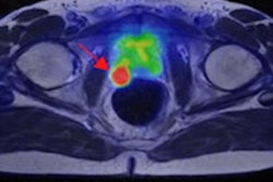
An experimental zirconium-89-based (Zr-89) PET radiotracer can detect tumors missed by existing tracers in patients whose cancer has returned after surgery or radiotherapy, according to a study published in the 1 April issue of the Journal of Nuclear Medicine.
German researchers tested a novel Zr-89-based prostate-specific membrane antigen (PSMA) tracer for the first time in a small group of patients. They found it detected PSMA-positive lesions in more than half of those who had previous negative scans with established tracers. Moreover, the scans enabled treatments for these patients, the authors wrote.
"Zr-89 PSMA-DFO might be used in men with [biochemical recurrence] after a PET/CT scan with established PSMA tracers was read as negative," wrote first author Dr. Felix Dietlein, PhD, of the University Hospital of Cologne.
PSMA is a protein expressed on prostate cancer cells, and radiotracers designed to bind to PSMA can indicate on imaging whether cancer has relapsed after patients undergo treatment. Gallium-68 (Ga-68) PSMA-11 has proven effective for this purpose, and it was approved for use in the U.S. in December 2020.
Nevertheless, PSMA-PET scans fail to identify the locations of tumor lesions in approximately 20% of patients with biochemical recurrence, according to the authors.
In this study, Dietlein and colleagues studied the use of Zr-89 PSMA-DFO for PET imaging in 14 prostate cancer patients diagnosed with biochemical recurrence based on rising PSA serum levels. The patients had previous negative PET scan results using Ga-68 PSMA-11 and F-18 JK-PSMA-7, an experimental tracer that has shown improved performance over Ga-68 PSMA-11 in preliminary studies.
Within five weeks after the negative scans, the researchers performed a second PSMA-PET scan (Biograph mCT, Siemens Healthineers) on the patients using Zr-89 PSMA-DFO. Most patients (11/14) had undergone prostatectomy followed by salvage radiotherapy.
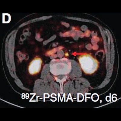
 Ga-68 PSMA-11 PET with maximum-intensity-projection (MIP) (A) and PET/low-dose CT fusion images (C), and Zr-89 PSMA-DFO PET six days after injection with MIP (B) and PET/low-dose CT fusion images (D) of a patient with biochemical recurrence after prostatectomy and stereotactic radiation therapy. Images are highly suggestive of PSMA-positive lymph node metastases supraclavicular left and retroperitoneal left, visible with Zr-89 PSMA-DFO (red arrows in B and D). Lymph nodes were PSMA-negative with Ga-68 PSMA-11 (yellow arrow in C). Patient underwent SRT and temporary ADT and PSA level dropped to 0.17 ng/mL. Image courtesy of Journal of Nuclear Medicine.
Zr-89 PSMA DFO detected 15 PSMA-positive lesions in eight of 14 patients who had a PET-negative reading of their initial PET scans with existing tracers, according to the findings. In these eight patients, the new scans revealed localized recurrence of disease (3/8), metastases in lymph nodes (3/8), or lesions at distant sites (2/8).
Moreover, on the basis of these results, patients received lesion-targeted radiotherapies (5/8), androgen deprivation therapies (2/8), or no therapy (1/8).
"Zr-89 PSMA-DFO may therefore offer a benefit to patients with weak PSMA positivity, in whom the localization of recurrent tumor lesions has proved challenging using existing PSMA tracers," the authors wrote.
Ultimately, existing PSMA tracers are limited by the short half-life of their radioactive labels (1.1 hours for Ga-68, for instance), which means that PET images must be acquired within three hours of injection. Yet experimental data suggest that internalization of PSMA ligands gradually increases over time, according to the authors.
Thus, since Zr-89 has a half-life of 77 hours, it may hold an inherent advantage over existing tracers, they suggested.
"If PET/CT images could be acquired much later, such as days after injection, even prostate cancer lesions with weak PSMA expression might become detectable on PSMA PET scans," they wrote.
In this study, PET scans with existing tracers were acquired one hour after injections with Ga-68 PSMA-11 and two hours after injections with F-18 JK-PSMA-7, while Zr-89 PSMA-DFO PET/CT images were acquired two and three days after tracer injection.
The authors noted this was a first-in-human study and that the data provides at least a solid rationale to further evaluate the performance of Zr-89 PSMA-DFO in prospective clinical trials.
"Larger clinical cohorts will be required to characterize and confirm the clinical benefits of 89Zr-PSMA-DFO," Dietlein and colleagues concluded.