
LIVERPOOL -- Be aware that tattoo pigments can mimic pathology and introduce artifacts on x-rays, causing potential errors. That's the key message of new research being presented at this week's U.K. Imaging and Oncology (UKIO) congress.
"Tattoo pigment composition significantly influences radiographic appearance and image quality, with higher atomic number and density pigments producing greater radiopacity and detectability," noted first author Maxwell Allen, senior author Ruwan Wanni Arachchige, PhD, and colleagues from the University of Exeter. "Yellow and white pigments (titanium dioxide) showed the greatest detectability, followed by blue and green (copper-based), while red (iron-based) and black (carbon-based) showed the lowest detectability."
 Growing social acceptance of tattoos means more patients now present with them.Courtesy of Julian and Courtney Astbury
In x-rays, these elements increase attenuation through photon-electron interactions, influenced by atomic number and density, they explained. As tattoo pigments vary in metallic composition, they may alter image quality. In mammography studies, normally radiotransparent areas can appear as radio-opaque calcifications, and such misrepresentation can lead to diagnostic errors, delayed treatment, and poorer patient outcomes.
The researchers' main goal was to investigate how variations in tattoo pigment composition affect radiotransparency in x-ray imaging. They sought to quantify the radiotransparency between tattoo pigments using contrast-to-noise ratio (CNR) analysis, to identify and characterize radiographic artifacts associated with different tattoo pigments, and to interpret how these findings may influence image quality.
They used a mixed-method phantom experimental study to assess the effect of tattoo pigment composition on radiotransparency. Pigment type was the independent variable; CNR and visual artifacts were dependent measures. Pilot testing standardized tattoo area (2 × 2 cm), ink volume (0.5 mL), and established controls (no-interference, needle only, pooled ink).
"Porcine skin was selected due to its similarity to human tissue," Allen and colleagues told delegates. "26 samples were prepared: 18 tattooed (six pigments in triplicate), six pooled-ink controls, and two additional controls. Pigments (black, white, blue, green, yellow, red) were selected based on elemental composition. Samples were shaved, cut (7 × 7 cm) and tattooed using a controlled depth (0.2 cm) and voltage (6.5 V), selected for adequate ink deposition while minimizing full-thickness penetration of the porcine skin."
The researchers acquired radiographs using digital radiography (41 KVp, 1.1 mAs, 100 cm SID). They used nine 45 × 45-pixel regions of interest (three tattooed, three adjacent tissues, three background) to calculate sample and mean pigment-group CNR.
Key findings
Mean CNR was highest for yellow (1.426), followed by white (0.75), blue (0.661), green (0.449), black (0.331); it was lowest for red (0.306). Pooled-ink controls generally showed higher CNR than tattooed samples, except black. The needle-only control had the lowest CNR (–0.550).
"One-way ANOVA demonstrated a major difference in mean CNR between pigments," the researchers added. "One-sample t-tests showed only yellow and white had CNR values significantly greater than zero, indicating radiographic detectability. Blue, green, black, and red were not significantly different."
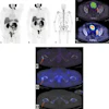
 Representative radiographs of black (left) and yellow (right) tattoo pigment samples demonstrating standardized regions of interest (ROIs) used for contrast-to-noise ratio (CNR) analysis. Presented by the Exeter team at UKIO 2026 and ECR 2026 (see https://epos.myesr.org/poster/esr/ecr2026/C-29619).Allen et al
Yellow, white, blue, green, and red tattooed samples and pooled-ink controls demonstrated a metallic artifact, appearing radio-opaque. Black tattooed samples and the needle-only control showed a tissue-disruption artifact, appearing as radiolucent regions due to needle penetration. The black pooled-ink control showed slight localized opacity due to additional pooled surface ink, while the no-interference control showed no visible artifact.
 How precisely do black tattoos affect x-rays? The researchers tackled this tricky question. Courtesy of Julian and Courtney Astbury
In 2025, the European Academy of Dermatology and Venereology (EADV) published guidelines about the impact of tattoos on imaging.
"In most cases, tattoos do not interfere with x-ray imaging," EADV said. "On very rare occasions, tattoos might appear as shadows or artifacts on the image. Informing the technician helps avoid confusion during interpretation."
Tattoos on the breast or chest wall may show up on a mammogram, according to the guidelines. "Pigments from tattoo ink that have migrated in a local axillary or intramammary lymph node may be visible on mammograms and can be mistaken for calcifications."
UKIO takes place from 8 to 10 June. More details on the Exeter group's study can be found here. The coauthors were Liam Byrne, Gkinovefa Tzogka, Emily Williams, and Pradeepa Ruwan Wanni Arachchige.


![A normal mammogram confirmed by three-year radiologic follow-up illustrates reader-marked regions of interest (ROIs) during (A) unaided (round 1) and (B) artificial intelligence (AI)–assisted (round 2) reading. Each colored dot represents an ROI for recall by a human reader. Readers could mark more than one ROI per case, represented by multiple dots of the same color. During AI-assisted reading, the AI system displayed three visible prompts: two with suspicion of malignancy scores of 35% (left mediolateral oblique [L MLO] and craniocaudal [L CC]) and one with a suspicion of malignancy score of 10% (right craniocaudal [R CC]), shown as polygonal overlays. Without AI, six of 10 readers (60%) marked a false-positive ROI. With AI assistance, this fell to two of 10 (20%). R MLO = right mediolateral oblique.](https://img.auntminnieeurope.com/mindful/smg/workspaces/default/uploads/2026/07/2026-07-14-radiology-mammogram-ai-auto-bias.H0bYO8QlWs.jpg?auto=format%2Ccompress&dpr=2&fit=crop&h=167&q=70&w=250)
