AI scoring could pick out early signs of breast cancer up to six years before a diagnosis, according to research published June 9 in Radiology.
Cancer prediction scores issued by three AI-CAD systems were higher, on average, for women who were eventually diagnosed with breast cancer compared to scores in women who remained cancer-free, wrote a team led by Fredrik Strand, MD, PhD, from Karolinska University Hospital in Stockholm, Sweden.
“There is a lot of cancer that goes undetected during screening, and we’re particularly interested in how AI can help with this,” Strand told AuntMinnie. “Six to eight years before diagnosis, you could see that the AI cancer detection scores on average started increasing … with a sharper increase toward the last two to three years.”
AI-CAD systems continue to be explored in screening mammography. One study published in January showed that AI-supported screening mammography can find more cancers while reducing interval cancer diagnosis. Another study published in March showed the safety and efficacy of an AI-supported screening approach that excludes low-risk mammograms from radiologist reading.
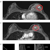
 Screening mammograms and AI score changes are depicted over time in two individuals with screen-detected cancer. Full-field digital mammograms show craniocaudal
(top) and mediolateral (bottom) views of the left and right breast. The description below each mammogram indicates whether AI scores from three AI-based computer-aided detection systems (AI-1, AI-2, and AI-3) were above or below the 90th percentile. (A) Mammograms from three screening time points (in 2009, 2011, and 2014) in an individual who was diagnosed in February 2014 (at age 73 years) with a left-sided 25-mm grade 2 invasive breast carcinoma, no special type (arrow). Mammographic breast density was classified as BI-RADS B on all three mammograms. (B) Mammograms from four screening time points (in 2009, 2011, 2013, and 2015) in an individual who was diagnosed in October 2015 (at age 65 years) with a left-sided 45-mm grade 2 lobular carcinoma (arrow). Mammographic breast density was classified as BI-RADS C on all four mammograms. Screen-detected cancers were defined as those diagnosed within 90 days for individuals who were recalled at screening (*).RSNA
Still, Strand and colleagues noted that AI’s role in early cancer detection needs further study. The team researched how AI scores in women diagnosed with breast cancer may increase up to 10 years before their diagnosis, compared with those who stay cancer-free.
The retrospective study used the Validation of Artificial Intelligence for Breast Imaging (VAI-B) database. This database covers four regions in Sweden, with data gathered from 2008 to 2019. The team included all mammography exams from each woman, with a maximum of 10 years before diagnosis in those who developed cancer.
The researchers used three commercial AI systems: Vara version 2.1, Vara AI (AI-1); Lunit Insight MMG version 1.1.7.2, Lunit (AI-2); and MammoScreen version 2.1.0, Therapixel (AI-3). The systems provided raw exam-level AI scores, which were converted to rank-order percentile AI scores.
Fredrik Strand, MD, PhD, explains what his team's findings could mean for AI-assisted breast imaging and breast cancer care.
Final analysis included 31,394 women with 88,963 mammography exams. Of these, 12,082 (38.5%) were diagnosed with breast cancer.
At 90% specificity, the AI-CAD systems flagged nearly one in five cancers at six years before diagnosis. They also significantly outperformed mammographic density as a predictor of breast cancer, which had an area under the receiver operating characteristic curve (AUC) of 0.57.
Performance of AI systems in predicting breast cancer | |||
Measure | AI-1 | AI-2 | AI-3 |
10 years before diagnosis | 12.7% | 13.8% | 17% |
Six years before diagnosis | 19% | 19.6% | 19.7% |
Four years before diagnosis | 24.2% | 23.3% | 25.2% |
For all time points combined, excluding screen detection, the area under the receiver operating characteristic curve (AUC) ranged from 0.63 to 0.67 for the AI-CAD systems. These all outperformed mammographic density (AUC = 0.57) as the predictor (all p < 0.001).
The researchers highlighted the potential of AI, possibly together with image-based risk factors, could make way for an early alert for supplemental imaging.
Strand told AuntMinnie that the team is interested in combining image-based information with genetic information to better stratify breast cancer risk in women.
Read the full study here.


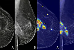


![A normal mammogram confirmed by three-year radiologic follow-up illustrates reader-marked regions of interest (ROIs) during (A) unaided (round 1) and (B) artificial intelligence (AI)–assisted (round 2) reading. Each colored dot represents an ROI for recall by a human reader. Readers could mark more than one ROI per case, represented by multiple dots of the same color. During AI-assisted reading, the AI system displayed three visible prompts: two with suspicion of malignancy scores of 35% (left mediolateral oblique [L MLO] and craniocaudal [L CC]) and one with a suspicion of malignancy score of 10% (right craniocaudal [R CC]), shown as polygonal overlays. Without AI, six of 10 readers (60%) marked a false-positive ROI. With AI assistance, this fell to two of 10 (20%). R MLO = right mediolateral oblique.](https://img.auntminnieeurope.com/mindful/smg/workspaces/default/uploads/2026/07/2026-07-14-radiology-mammogram-ai-auto-bias.H0bYO8QlWs.jpg?auto=format%2Ccompress&dpr=2&fit=crop&h=167&q=70&w=250)


