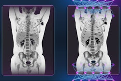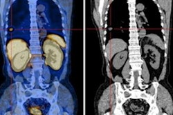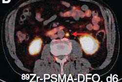
A PET/CT radiomics model based on imaging with a gallium-68 (Ga-68) PSMA-11 radiotracer could help clinicians diagnose primary prostate cancer, according to a study by Chinese researchers published September 30 in EJNMMI Research.
A group at Nanjing Medical University in Nanjing developed an algorithm based on features from Ga-68 PSMA-11 PET/CT scans of patients diagnosed with proven prostate cancer or benign prostate disease. In testing, the model outperformed visual assessments by nuclear medicine radiologists.
"Radiomics analysis based on 68Ga-PSMA-11 PET may non-invasively predict intraprostatic lesions in patients with [prostate cancer]," wrote corresponding author Dr. Feng Wang, PhD, a professor of nuclear medicine at Nanjing Medical University, and colleagues.
Prostate cancer is the second leading cause of cancer-related death in men worldwide, with rates increasing. Currently, image-guided biopsy is the standard method for making a definitive diagnosis, yet biopsies can fail to detect some cases and have notable side effects, including bleeding, pain, and infection, according to the authors.
The discovery that prostate-specific membrane antigen (PSMA) protein is overexpressed on the surface of prostate cancer cells led to the development of PSMA ligands that bind to the protein. When these ligands are combined with isotopes such as gallium-68 (Ga-68), the resulting radiotracers allow researchers to locate these tumors noninvasively on PET imaging.
Ga-68 PSMA-11 was approved in the U.S. in December 2020 for imaging in patients with suspected metastases, as well as in patients with suspected recurrence based on elevated prostate-specific antigen (PSA) levels. Research since suggests that the radiotracer is also highly accurate for diagnosing primary tumors in these patients.
Still, significant numbers of intraprostatic lesions can be missed by visual PET image interpretation due to their small size or configuration. In this study, the researchers hypothesized that a radiomics approach based on quantitative measures of PSMA expression may fill this diagnostic gap.
The group culled data from 125 consecutive patients diagnosed with prostate cancer or benign prostate disease (BPD) who underwent Ga-68 PSMA-11 PET/CT scans (uMI 780, United Imaging) at Nanjing First Hospital between February 2019 and May 2021. Patients had received no antitumor treatment before the PET/CT exams.
The researchers extracted 944 prostate cancer features from the images, including shape features, first-order intensity statistics features, and texture features. Then, they narrowed the field to nine features with the least redundancy and greatest correlation with the target tumor, which they used to train the model.
In a subsequent test set (25 patients with prostate cancer and 13 with BPD), the model achieved good predictive performance, with a sensitivity of 0.84, a specificity of 0.77, and a positive predictive value (PPV) and negative predictive value (NPV) of 0.88 and 0.71, according to the findings.
The area under the curve (AUC) value for the model for differentiating prostate histopathology was 0.85, compared with an AUC of 0.63 for visual assessments by trained nuclear medicine physicians who also analyzed the test set. In addition, the radiomics model achieved greater sensitivity, specificity, PPV, and NPV than those of the readers, the researchers wrote.
| Performance of readers vs. radiomics model for detecting prostate cancer on Ga-68 PSMA-11 PET/CT | ||
| Readers | Radiomics model | |
| Sensitivity | 0.74 | 0.84 |
| Specificity | 0.55 | 0.77 |
| PPV | 0.80 | 0.88 |
| NPV | 0.46 | 0.71 |
"The model was successfully validated in an independent test set and outperformed visual assessments by nuclear medicine radiologists," the authors wrote.
They noted limitations of the study, namely its single-center design and relatively small sample size, and ultimately called for a unified standard for multicenter studies to establish and test multicenter data using radiomics methods to improve the robustness of models.
Nonetheless, visual assessment of primary prostate cancer on PET/CT based on experience remains challenging. Although PSMA protein is highly expressed on the cell surface of prostate cancer cells, it is also expressed in benign pathologies such as BPD, which can further confound diagnoses, the researchers wrote.
"In this study, the radiomics model detected prostate cancer based solely on a numeric feature set from high-dimensional medical imaging data, regardless of the clinical situation," Wang and colleagues concluded.