Advanced imaging techniques like perfusion and diffusion imaging have successfully entered routine clinical practice, but broader adoption of quantitative MRI biomarkers requires standardized acquisition protocols, harmonized postprocessing, biological validation, and prospective evidence that these techniques actually change clinical decisions.
- Established techniques: Perfusion imaging and diffusion imaging with ADC maps are already integrated into daily neuroradiology practice for tumor and stroke imaging.
- Emerging methods: APT-CEST is transitioning to routine use through product sequences, while relaxometry mapping and sodium imaging remain primarily research tools.
- Three translation barriers: Acquisition harmonization across MRI manufacturers, standardized postprocessing and analysis, and prospective clinical validation showing biomarkers change treatment decisions.
- Standardization approach: Multiparametric quantitative imaging protocols developed across 13 German centers demonstrate the need for reproducible, cross-vendor eight-minute protocols that integrate with clinical workflow.
- AI advancement: Deep-learning reconstruction and foundation models are moving from proof-of-concept toward genuine clinical applications in lesion detection and analysis.
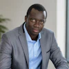
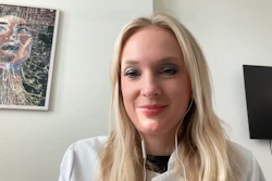
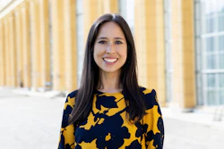
 neuroRAD's congress presidency: Dr. Roland Schwab, Magdeburg, Dr. Katharina Wenger-Alakmeh, Frankfurt am Main and Dr. Daniel Kaiser, Dresden.linkedin
AuntMinnieEurope: Which studies in this year’s abstract program look most likely to make a difference in clinical practice?
Katharina Wenger-Alakmeh: What convinced me this year was that there were many studies addressing very concrete clinical questions. The translational aspect really came through across the abstract program.
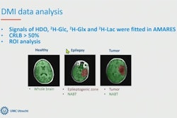
On the advanced MRI side, there was a lot on techniques ranging from CEST and diffusion imaging to multiparametric quantitative imaging. Much of this work focused on tumors, including tumor characterization, progression and the delineation of tumor margins.
That is becoming increasingly important, particularly with infiltrative tumor types, where we want to characterize the tumor margins better and make treatment planning more precise.
There was also interesting work beyond tumor imaging. One study from Freiburg looked at whether deep-learning reconstruction could increase sensitivity for very small lesions compared with standard imaging. We are also increasingly seeing work on foundation models, with several interesting studies from Bonn.
AME: Advanced imaging produces promising biomarkers, but many never reach routine care. Where does translation currently break down?
Wenger-Alakmeh: There are essentially three steps.
The first is acquisition. Particularly in MRI, we have different manufacturers and technologies that are not yet fully harmonized. This applies especially to multiparametric quantitative techniques, but also to spectroscopy if we want to quantify it.
One approach is to investigate this much more systematically. We have done that with multiparametric quantitative imaging in a project that started in Frankfurt together with 12 other centers in Germany.
We said that we need a reproducible, cross-vendor eight-minute protocol that can run in addition to clinical routine.
Then the other points come in: we also need harmonized postprocessing and analysis. Ideally, all the data should be standardized on a common platform, with as much of the analysis automated as possible.
Another point is biological validation. And ultimately we need to show prospectively that these biomarkers are robust enough to use and that they actually change clinical decisions.
AME: Which quantitative MRI techniques are already ready for routine use, and which are still mainly research tools?
Wenger-Alakmeh: For me, perfusion imaging has arrived in daily practice, particularly in tumor imaging. Diffusion imaging with ADC maps is of course also established, both in stroke and in tumor imaging.
In spectroscopy, proton spectroscopy is currently useful for selected questions. But, as I said, quantification is still not sufficiently comparable across manufacturers for broader use.
If we take quantitative imaging in a wider sense, there is also automated volumetry and lesion quantification, particularly in tumors, multiple sclerosis and neurodegenerative diseases.
APT-CEST is at an interesting point. Product sequences are now available and are increasingly being used. Because it is available as a product sequence, I think it will find an easier path into routine practice.
If we look at relaxometry mapping, sodium imaging and multinuclear imaging, however, we are still predominantly in the research domain, although we are moving forward slowly.
AME: AI is moving from proof-of-concept studies toward clinical questions. Which developments at neuroRAD suggest it is becoming genuinely useful?
Wenger-Alakmeh: There was interesting work on deep-learning reconstruction, including the study from Freiburg looking at sensitivity for very small lesions compared with standard imaging.
And increasingly, we are seeing studies on foundation models. There were several interesting contributions on that topic as well.
AME: If European radiologists take one message home from neuroRAD 2026, what should it be?
Wenger-Alakmeh: I think I would choose the generational change and how we have tried to make that concrete. We have a three-person presidency, and for the different formats we pair younger chairs with experienced mentors.
That creates new connections as well. It means having someone you can contact later when you have a question, someone who can help you and give you insight.
I also think it is important to create formats that specifically consider the needs of women in neuroradiology, or in other specialties. Many of these topics overlap with those affecting the younger generation more broadly, but there are also issues that may need to be discussed in a more protected space.