PET imaging with an experimental radiotracer called F-18 Florzolotau shows promise for the diagnosis of early progressive supranuclear palsy (PSP), a team of researchers has found.
Investigators from Fudan University in Shanghai used the tracer to identify brain PET imaging patterns of tau protein in a large group of PSP patients.
"[Our] data indicate that F-18 Florzolotau PET imaging holds promise for tracking the accumulation of tau pathology in the living brain of patients with [progressive supranuclear palsy]," wrote lead author Dr. Feng-Tao Liu, PhD, and colleagues. The study was published on 11 January in the European Journal of Nuclear Medicine and Molecular Imaging.
PSP is a rare brain disorder that causes problems with movement, walking and balance, and eye movement. There are no current treatments for the disorder, nor are there definitive imaging approaches for detecting it. Until now, MRI has been considered the most useful diagnostic tool for the disease, according to the authors.
However, transgenic mice and human postmortem studies have suggested that brain tau pathology -- specifically, the presence of the four-repeat (4R) form of tau protein -- plays a significant role in PSP and that PET can illuminate this pathology.
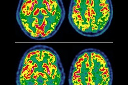
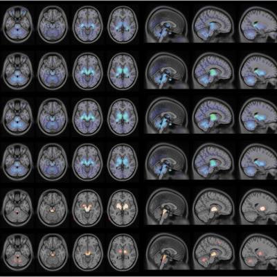
 F-18 Florzolotau PET images of patients with progressive supranuclear palsy. The images illustrate F-18 Florzolotau uptake in the healthy control (HC) group (panel A), entire patient cohort (panel B), as well as in the PSP-Richardson's syndrome (RS) (panel C) and PSP-non-RS (panel D) groups. Compared with the HC group, patients with PSP showed significantly increased binding mainly in the pallido-nigro-luysian axis (panel E). While the patterns of tracer accumulation were similar in the two patient groups, the binding intensity was markedly lower in patients with PSP-non-RS (panel F). The color bars in panels A-D indicate standardized uptake value ratios (SUVR), whereas the color bar in panels E-F denote threshold values from the voxel-wise comparisons. Image and caption courtesy of the European Journal of Nuclear Medicine and Molecular Imaging through CC BY 4.0.
Liu's team sought to confirm findings from these preclinical studies in a large group of patients using PET imaging with F-18 Florzolotau, a tracer originally developed in 2020 that has shown promise for detecting hallmark tau pathology in Alzheimer's disease. To do so, the investigators recruited 148 consecutive patients with PSP to undergo F-18 Florzolotau PET imaging between May 2019 and January 2022. Twenty subjects with a negative history for neurological or psychiatric disorders were included in a control group for comparison. The researchers tracked 18 subcortical regions of interest and eight cortical regions of interest.
Based on standard radiotracer uptake measurements, the distribution patterns of F-18 Florzolotau uptake in the regions of interest showed a striking similarity to those reported in postmortem studies, with the binding intensity of the tracer to its tau target markedly higher in PSP-/Richardson's syndrome, the classic type of PSP. The tracer also detected tau accumulation at earlier stages of the disorder compared with postmortem immunostaining findings, they noted.
The study findings warrant further research, according to the authors.
"Patients with PSP should be followed longitudinally in relation to the distribution patterns to evaluate whether this classification may inform prognosis," Liu and colleagues concluded.