Of all the medical imaging modalities, perhaps none has demonstrated its value for diagnosing patients with COVID-19 more than CT. But how exactly should CT be used in clinical practice?
Despite CT's demonstrated value, there is some controversy around the performance of CT for COVID-19 diagnosis, said Dr. Marie-Pierre Revel, PhD, professor of radiology and head of cardiothoracic imaging at Paris Descartes University's Cochin Hospital.
The reason is that the standard of reference, reverse transcription polymerase chain reaction (RT-PCR) testing, is itself imperfect, she said. It is not unusual for clinicians to diagnose COVID-19 infections on CT scans while RT-PCR tests are negative -- sometimes more than once.
 Dr. Marie-Pierre Revel, PhD, of Paris Descartes University's Cochin Hospital in Paris, France, presenting on 2 March at ECR 2022 Overture.
"These are what I would call 'false false-negatives,' " Revel noted during a special session at the recent ECR 2022 Overture meeting.
With this backdrop, Revel led a series of talks by experts who recommended effective CT imaging approaches for COVID-19 patients in emergency settings.
Severity scoring systems
Dr. Ivana Blazic, PhD, MRI section head in Clinical Hospital Center Zenum in Belgrade, Serbia, discussed the nuts and bolts of scoring systems for CT imaging findings in patients.
"The use of severity scoring systems as a part of the standard report of chest CT for COVID-19 patients is highly recommended," Blazic said.
CO-RADS, a five-point scheme developed by the Dutch Radiological Society, as well as the RSNA's chest CT classification system, are comparable for reporting findings attributable to COVID-19, with each providing standardized language and sensitivities of about 75%, she said.
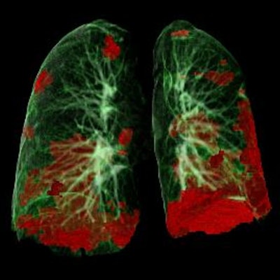
A comparison last year among four scoring systems -- chest CT score, total severity score (TSS), CT severity score (CT-SS), and three-level chest CT severity score -- showed excellent interobserver agreement between the systems and no significant differences in receiver operating characteristic (ROC) curves. Yet evidence points to chest CT score and TSS, Blazic said.
"Chest CT scoring system and total severity score achieved the highest specificity and are least time consuming," she said, and added the use of chest CT score is standard now at her hospital in Belgrade.
Ultimately, the use of severity scores are associated with better clinical outcomes for patients, Blazic concluded.
The 'great radiological mimicker'
Differential diagnosis on CT in COVID-19 patients is difficult, primarily because CT findings have low specificity due to their overlap with a number of other conditions, according to Dr. Anna Rita Larici of the Catholic University of Sacro Cuore in Rome, Italy.
"Since the beginning of our experience, we've learned COVID-19 pneumonia should be considered a great radiological mimicker," she said.
Other conditions to consider include diffuse alveolar damage, acute respiratory distress syndrome (ARDS), alveolar proteinosis, and pneumocystis jiroveci pneumonia. However, CT-related findings are significantly more common in COVID-19 compared with other viral respiratory illnesses, Larici said.
 Dr. Anna Rita Larici of the Catholic University of Sacro Cuore in Rome, Italy, presenting on 2 March at ECR 2022 Overture.
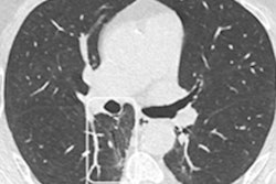
Primary CT features in favor of COVID-19 pneumonia include ground-glass opacification (GGO) -- patchy, peripheral, bilateral, and basal predominant distributions -- and enlarged pulmonary vessels, while features against COVID-19 pneumonia include the following:
- Lack of GGO
- Lobar or nonsegmental consolidation
- Cavitation/abscesses
- Tree-in-bud opacities
- Lymphadenopathy
- Pleural effusion
- Pericardial effusion
"The combination of CT features with clinical data, history, and lab and microbiology in the multidisciplinary team context can substantially improve the accuracy of diagnosis and patient management," Larici said.
Which modality is best?
Dr. Luis Gorospe Sarasua of Ramón y Cajal University Hospital in Madrid, Spain, said CT is an excellent modality in emergency settings for evaluating the extent of COVID-19 disease, detecting complications, and for indicating differential diagnoses. However, chest x-rays have proven very useful and may be recommended in cases where resources are limited.
Most radiological societies recommend the use of CT under the following three conditions:
- In COVID-19-confirmed patients with disease progression, especially if complications are suspected
- In patients in whom there are clinical, lab, or chest x-ray discrepancies -- for instance, in severely ill suspected COVID-19 patients with normal chest x-rays and negative or unavailable PCR
- In suspected COVID-19 patients in whom quick decisions need to be made regarding admission to the intensive care unit
"There is no role for imaging in mild cases when resources are limited," Sarasua said.
If a patient's symptoms are mild, management strategies should start with an RT-PCR test. If positive, CT imaging should only be indicated in patients at risk for progression, Sarasua said.
If symptoms are moderate to severe, imaging is indicated no matter the results of the COVID-19 test, he said. The imaging modality used depends on availability and local protocols. CT is suggested if patients experience progression at the emergency department, while chest x-rays may be preferred if resources are limited, he said.
"No matter what modality is used, consistent and prompt reporting using categorization systems and scoring systems is strongly recommended," Sarasua concluded.
Revel concluded the ECR session with a comment on CT she said everybody should agree on -- the modality has proven the efficacy of vaccinations. In a recent study, the percentage of CT scans without pneumonia was 22% in unvaccinated patients, compared with 59% in vaccinated patients.
Perhaps spreading that simple message can convince people still resistant to vaccinations to get them, she said, "because seeing is believing."