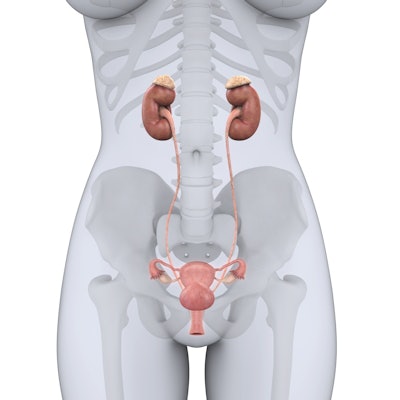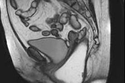
MRI can supply vital information about the anatomy and pathology of the female urethra, and it can guide clinical management and provide a road map for surgical procedures, according to new analysis by European genitourinary radiologists.
"MRI is a useful imaging tool for the primary assessment of the female urethra for both benign and malignant pathology," Dr. Roxana Pintican, from Iuliu Hatieganu University of Medicine and Pharmacy, Cluj-Napoca, Romania, and colleagues at the University of Cambridge, U.K., reported at RSNA 2021. "MRI is able to characterize incidentally depicted urethral lesions."
The use of pelvic MRI has rapidly increased over the last decade, for example in the preoperative imaging of patients with pelvic cancers, such as endometrial and cervical cancer. Therefore, incidental urethral pathology is more commonly detected, increasing the need for accurate image interpretation and diagnosis, they noted in an e-poster presentation.
The female urethra is well delineated on pelvic MRI due to the excellent soft tissue and spatial resolution of MRI. In addition, MRI has an evolving role in the primary assessment of the female urethra for both benign and malignant pathology, the researchers continued.
Retrograde urethrography and voiding cystourethrography are typically the primary imaging modalities of choice in the male urethra, but these approaches provide less information concerning the female urethra. Pintican and her colleagues' aim was to highlight female urethral pathology on MRI, both as a primary tool to investigate suspected urethral pathology and for the incidentally depicted pathology, secondary to the increasing usage of pelvic MRI in oncology patients.
MRI protocols
Currently there are no studies in the literature about the usefulness of using diffusion-weighted imaging (DWI) or contrast enhancement, the authors noted.
"Our experience has shown that gadolinium is very useful to evaluate urethral integrity and helps to identify urethral mesh erosion," they pointed out. "Furthermore, we use DWI/ADC (apparent diffusion coefficient) in all cancer patients for a better tumor delineation and to increase our diagnostic certainty."
 Recommended urethral MRI protocol. T1w = T1-weighted, FS = fat-suppressed, FOV = field-of-view, TRUFI = TrueFISP or fast imaging with steady precession.
To evaluate a case, they recommend the following steps:
- Check the anatomy -- is everything in place?
- Follow the urethra from top to bottom -- localize the abnormality (upper, mid, lower urethra)
- Is there a cystic or solid abnormality?
- Is there a clear communication/contiguity between urethra and abnormality?
- Do not forget the rest of the pelvis. Sometimes the primary diagnosis arises from another organ (e.g., cancer in cervix).
Embryology is fundamental to understanding the complex female malformations, combining mesonephric, Mullerian, cloacal, and urogenital sinus anomalies. Their overall prevalence is estimated to be 3%-6%, being responsible for infertility and clinical symptoms and having a severe impact on quality of life, especially in young women, the authors stated.
Isolated female urethra malformations are extremely rare, and it is more common for additional structures to be involved such as the bladder (bladder-sphincter dysfunction) or vagina (congenital urethrovaginal fistula), they added.


