Dear AuntMinnieEurope Member,
Spare a thought for the 15 radiology trainees at Westmead Hospital in Sydney. Just when they require stability and expert supervision, they face an uncertain future after the radiology department lost its training accreditation.
This might happen elsewhere in the months and years ahead. Training schemes across the globe are under intense pressure as a result of the pandemic, and the increasing emphasis on radiology workload and volume is creating extra tension and difficulties.
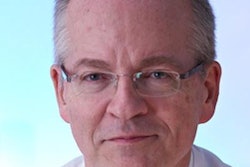
Further evidence of Dr. László Tabár's remarkable longevity and vision came to light this week, when we posted an interview with him about breast artificial intelligence (AI). He acknowledges the great potential of AI, but he also has some reservations. Find out more in the Women's Imaging Community.
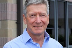
Like many of us, Dr. Giles Maskell has loved the action from the Tokyo Olympics, and he's wondering whether a radiology Olympics might be feasible. In a light-hearted column, he proposes several possible events, including speed reporting, cherry-picking, and the surgical conversation.
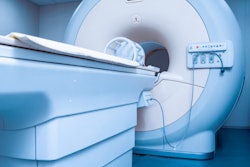
After three exhausting weeks in the polyclinic, chief radiologist Dr. Yukihisa Saida and his team must be enjoying some rest and relaxation before the start of the Paralympics on 24 August. Soon after Sunday's closing ceremony, he spoke to us again about the imaging of the Games. Learn more in the MRI Community.
Last but not least, a new study in the British Journal of Cancer caught our attention. Chinese authors have used deep learning to identify hepatocellular carcinoma on liver CT exams with a level of accuracy comparable to radiologists.