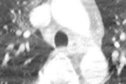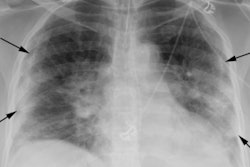
A survey of 50 radiology departments in 33 countries has highlighted significant variations in imaging procedures for COVID-19 patients. Conducted by the International Society of Radiology (ISR) and supported by the European Society of Radiology (ESR), the research was published online on 17 September by European Radiology.
 Dr. Ivana Blažić, PhD.
"All imaging departments involved in this survey reported the use of imaging in COVID-19 patients showing severe symptoms or who were critically ill," noted first author Dr. Ivana Blažić, PhD, MRI section head in Clinical Hospital Center Zemun in Belgrade, Serbia, and colleagues. "However, there is a wide variation in imaging modality type used for each clinical scenario."
The use of imaging is applied in line with existing guidelines and recommendations in 98% of facilities, with structured reporting recorded in 58% of institutions. No less than 83% centers reported a significant impact of the COVID-19 pandemic on the imaging department's routine activity, the authors added.
Suspected versus confirmed cases
The survey found that imaging is not usually performed in asymptomatic patients in around 69% of institutions, but it is used in suspected or confirmed patients with COVID-19 (89% and 94%, respectively), in COVID-19 patients showing severe symptoms or who were critically ill (100%), and at the end of confinement (60%). However, Blažić and colleagues found a wide variation in imaging modality type used for each clinical scenario.
In symptomatic patients with suspected COVID-19, the vast majority of imaging departments perform imaging (89%), of which 37% use chest x-ray (CXR), 34% chest CT, and 29% both CXR and CT. The main reasons given for imaging use were to receive faster results than molecular tests (51%) and ease of access (39%), while 5% of the institutions didn't have access to molecular tests.
 Map chart shows geographical variations in the use of imaging in patients with suspected COVID-19. Different answers from institutions in the same country are given in the side table; CXR = chest x-ray. Charts courtesy of European Radiology.
In patients with confirmed COVID-19, imaging is performed in the vast majority of imaging departments (94%), of which 22% use CXR, 28% chest CT, and 43% both CXR and CT. In some institutions, the combination of three modalities -- CXR, CT and lung ultrasound (LUS) or CT, LUS and MRI (4% and 2%) -- is used. The purpose of imaging use in patients with confirmed COVID-19 was the detection of lung disease (96%) or thrombotic complication (52%) related to COVID-19.
 Map chart shows geographical variations in the use of imaging in patients with confirmed COVID-19. Different answers from institutions in the same country are given in the side table.
In patients with COVID-19 exhibiting severe symptoms or critical illness, x-ray is performed in 10% and 33% of institutions, respectively. Chest CT is used in 31% and 19% of institutions, both CXR and CT in 49% and 23% of institutions, and three imaging modalities (CXR, CT, and LUS) in 6% and 13% of institutions, Blažić and colleagues reported.
Additionally, in patients with severe symptoms, the combination of chest CT and LUS was used in 4% of institutions, while in 13% of institutions, the combination of CXR and LUS was used in patients who were critically ill. The purpose of imaging use in patients showing severe symptoms or being critically ill was the detection of lung disease (98% and 88%, respectively) or thrombotic complication (73% and 54%, respectively) related to COVID-19.
Plethora of guidelines
The authors of the article -- who included Prof. Boris Brkljačić, chairman of the ESR Board of Directors, and Prof. Guy Frija, chair of EuroSafe Imaging -- also presented a summary of the recommendations of professional societies from countries with the largest proportions of COVID-19 patients.
"The guidelines on the use of imaging in COVID-19 differ at a smaller or larger extent depending on the country or region and the level of and access to healthcare in the respective country or area of the world and it seems that they also reflect the local situation of the COVID-19 pandemic," they noted.
Overall, the authors hope this survey will help to boost understanding of current heterogeneities in radiology practice and to identify needs and gaps in the organization and function of radiology departments worldwide in relation to the COVID-19 pandemic. They aim to assist with the development of an overall strategy for radiology department organization and imaging protocols in pandemic conditions.