X-ray imaging and advanced radiology techniques reveal hidden layers, alterations, and lost details in centuries-old paintings that are invisible to the naked eye, allowing art historians to see what master artists originally painted or later concealed beneath the surface.
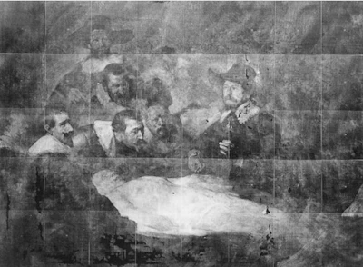
- X-ray imaging revealed that Rembrandt originally painted an amputated stump on the dissected arm in The Anatomy Lesson of Dr. Nicolaes Tulp, then added a hand later
- X-ray fluorescence scanning maps chemical elements like cobalt, copper, and lead to identify original pigments and detect colors that have faded or turned brown over centuries
- High-resolution microscopy discovered that tiny brown structures on a Rachel Ruysch flower painting were actual butterfly scales, not brushstrokes
- Technical art history merges imaging, chemistry, and microscopy to reveal how paintings were made, how they changed, and what remains hidden beneath visible surfaces
- Imaging allows researchers to examine altered layers without removing them, protecting original artwork from damage during the discovery process
Art that has been around for hundreds of years is familiar through its figures, colors and compositions. But radiology can peel back centuries of layers, revealing what has been hiding beneath the surface.
A scan can uncover in minutes what has remained hidden for centuries: a hand added later where there was once an amputated stump, clothes painted over a nude figure, colors that have faded into something completely different, or tiny marks thought to be brushstrokes that turn out to be scales from a real butterfly.
For radiologists, looking behind the surface is everyday work. At the opening of EUSOBI 2026 in Maastricht, that same way of seeing was turned on art.
Technical art history is a discipline that merges imaging, chemistry and microscopy to reveal how paintings were made, how they changed over time and what may still be hiding beneath the visible surface.
More than meets the eye

Technical art historian Paul van Laar, looking rather fittingly artsy himself and with a research background at the University of Cambridge, projected masterpiece after masterpiece onto the screen and then took the audience beneath the paint.
“What we see with the naked eye today might not be all that the painting has to offer,” he said.
One of the first examples was Rembrandt’s The Anatomy Lesson of Dr. Nicolaes Tulp. In the painting as it is known today, a hand lies at the end of the dissected man’s arm. But an x-ray revealed that Rembrandt had originally painted the arm ending in a stump and added the hand later.
 Artemisia Gentileschi, Allegory of Inclination (1816), in x-ray imaging.Courtesy of the Casa Buonarroti, Florence
Another example showed how drapery added to Artemisia Gentileschi’s Allegory of Inclination concealed the nude figure underneath. Imaging allowed researchers to look beyond the later layer without removing it and damaging the original painting.
While examining a flower painting by 17th-century artist Rachel Ruysch, van Laar noticed tiny brown structures on a butterfly. They appeared too small to be ordinary brushstrokes. Under high-resolution microscopy, they turned out to be actual butterfly scales.
- Recovering lost colors
Even the colors visible in paintings can tell a different story once imaging looks beneath them.
Using X-ray fluorescence scanning, researchers can map chemical elements such as cobalt, copper, iron, arsenic and lead across a painting. Because those elements are linked to particular pigments, the resulting maps can show what materials an artist used and where.
Even more, they can reveal colors that have vanished. Van Laar showed the example of smalt, a blue pigment made from finely ground cobalt-containing glass. Over centuries, the pigment can lose its blue appearance and turn brownish or gray. Yet cobalt can still be detected through X-ray fluorescence, allowing researchers to see where vivid blue may once have been present.
A painting that has looked brown for generations may therefore have looked dramatically different when it left the artist’s studio.
 Rachel Ruysch (North Netherlandish painter) 1664
The lecture was part of an opening ceremony that mixed science, art, and culture. EUSOBI President Dr. Michael Fuchsjäger led through the event with a playful succession of surprises, including a special ceremony honoring breast imaging expert Dr. Ruud Pijnappel, followed by a performance from Dutch pianist Jan Vayne.
 Presentation Technical art historian Paul van Laar
He also revealed another milestone for the society: EUSOBI was approaching 3,000 members. The auditorium was full and the industry area bustling.
While art made the opening, EUSOBI immediately turned back to breast imaging, with smarter screening, AI and personalized risk, contrast-enhanced mammography (CEM), minimally invasive interventions, sustainability, and emerging young researchers among the topics still to come.