While breast cancer imaging technology has advanced significantly with improved diagnostic tools like digital tomosynthesis and MRI, experts emphasize that successful screening and diagnosis depend equally on personalized approaches based on individual risk factors and clear patient communication throughout the diagnostic process.
- Breast cancer impact: Nearly 7,000 Austrian women diagnosed annually, representing 31% of new female cancer cases and approximately 19 new cases and 5 deaths daily
- Personalized screening shift: Modern approaches tailor screening based on age, breast density, genetic predisposition, and family history to improve detection while reducing unnecessary exams
- Mammography limitations: Detection rates range from 86-89% in fatty breasts to just 62-68% in extremely dense tissue, which itself increases cancer risk
- Complementary imaging tools: Digital breast tomosynthesis, ultrasound, and MRI enhance diagnostics when used strategically for individual patient needs
- Patient-centered care essential: Clear communication about findings, uncertainties, and next steps is critical, as abnormal imaging doesn't automatically indicate cancer
Advances in imaging technology and breast cancer research are improving diagnostics. Yet, as experts emphasized at an ESR event on 14 October, patients must remain at the center of every diagnostic decision.
Choosing the right examination
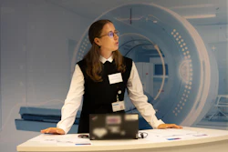
Prof. Minerva Becker, chairperson of the ESR Board of Directors and radiologist at Geneva University Hospitals, guided participants through the stages of breast cancer diagnosis, emphasizing that imaging modalities complement rather than replace one another.
Mammography remains central to breast cancer screening, particularly for detecting microcalcifications. However, its effectiveness depends partly on breast density. Becker presented detection estimates of 86%–89% in predominantly fatty breasts, compared with 62%–68% in extremely dense breast tissue.
Dense breast tissue can mask tumors on mammograms and is itself an independent risk factor for breast cancer. Becker therefore emphasized the importance of breast density awareness and considering individual risk factors when deciding whether additional imaging is needed.
Digital breast tomosynthesis can improve visualization by separating overlapping tissue structures, while ultrasound helps characterize suspicious lesions, distinguish cysts from solid masses, and guide biopsies.
Breast MRI can provide further information in selected patients, particularly when assessing disease extent, examining extremely dense breasts, or investigating elevated familial or genetic risk. The Dutch DENSE trial demonstrated that supplemental MRI in women with extremely dense breasts can reduce interval cancers. However, additional MRI findings do not necessarily indicate malignancy, she noted.
"Behind every image is a person"
"An abnormal imaging result does not automatically mean cancer," she said. Further examinations and, when indicated, a biopsy help establish a diagnosis and determine tumor characteristics. A biopsy itself does not imply that cancer is present.
Following a cancer diagnosis, imaging may also help assess disease spread. PET/CT combines metabolic and anatomical information but is generally reserved for selected indications, including certain higher-stage cancers or suspected metastatic disease.
Throughout the diagnostic process, Becker emphasized communication, outlining three questions: What do we know? What remains unclear? And how do we proceed? Explaining findings, acknowledging uncertainties, and deciding on the next steps together are essential to patient-centered care. "Behind every image is a person," she emphasized.
More than a diagnosis
The importance of patient support was also illustrated by Caroline Justich, founder of the ESR-supported initiative Be accepted and a representative of the ESR Patient Advisory Group.
Justich was diagnosed with metastatic breast cancer at age 39, exactly ten years before the Vienna event. She described the shock of receiving the diagnosis and how a radiologist helped her understand the findings and navigate the next steps. This experience inspired her to create Be accepted.
The initiative combines medical information with resources on psychological well-being, nutrition, physical activity, relationships, body image, and quality of life. Its octopus symbol represents intelligence, adaptability, and resourcefulness, reflecting the animal's ability to manage multiple challenges and escape seemingly hopeless situations.
With educational materials and digital resources, Be accepted aims to help patients understand their diagnosis, access reliable information, and take an active role in their care.
While advances in imaging and individualized risk assessment can improve clinical decisions, the event highlighted that patients also need clear information and guidance throughout their diagnostic and treatment journey.