When it comes to the risk of coronary artery disease (CAD), not all fat is created equal, say Hungarian researchers. They found that at least one type of visceral fat correlates with the presence of coronary artery disease.
 Dr. Zsofia Drobni from the Cardiovascular Imaging Research Group at Semmelweis University in Budapest.
Investigators from Semmelweis University examined healthy patients from the Burden of Atherosclerotic Plaque Study in Twins (BUDAPEST) to look for a relationship between fat in different regions of the abdomen and the presence of coronary artery disease using low-dose CT of the abdomen, as well as coronary CT angiography (CCTA).
They found that regions of visceral adipose tissue surrounding the heart were associated with the presence of coronary artery disease, while fat in other regions had no effect.
"Epicardial fat volume is an independent predictor of coronary artery disease," said Dr. Zsofia Drobni, from the Cardiovascular Imaging Research Group, in a presentation at ECR 2016. "This finding supports the hypothesis that epicardial fat promotes atherosclerosis development."
Visceral obesity a danger sign
The study measured epicardial fat volume (EFV), subcutaneous adipose tissue (SAT), and visceral adipose tissue (VAT), comparing each type to the presence of coronary artery disease as assessed by CCTA.
"Ectopic fat is defined as fat accumulation in tissues other than adipose tissue, which normally contains only small amounts of fat -- for example in the liver, in the heart, and in the pancreas," Drobni said. "Epicardial fat can be defined as fat found inside the pericardium."
CT can look for two main types of fat: subcutaneous fat, which accumulates under the skin, and fat that accumulates between the abdominal organs in the case of visceral obesity.
 Visceral adipose tissue (VAT) is known to be more dangerous to health than subcutaneous adipose tissue (SAT). In particular, visceral fat surrounding the heart raises the risk of coronary artery disease. All images courtesy of Dr. Zsofia Drobni.
The study included 195 healthy asymptomatic subjects with no history of coronary disease from the BUDAPEST study. All of them underwent low-dose noncontrast-enhanced CT, from which the study team quantified the epicardial fat volume. Every patient also underwent coronary CT angiography.
In the CCTA studies, readers assessed every coronary artery segment for the presence of atherosclerotic plaque, classifying the patients into groups with CAD and without CAD. For patients who were positive for coronary disease, they calculated the segment involvement score, representing the total number of arterial segments with plaque.
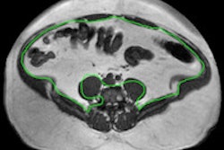
The researchers also measured epicardial fat volume from the CCTA data, while measuring the SAT and VAT fat types on a single CT slice acquired at the level of the L3/L4 vertebrae.
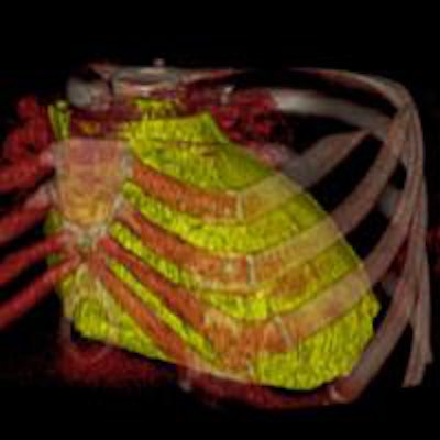
 3D reconstruction of epicardial fat volume. Epicardial fat (in yellow) is associated with a higher risk of coronary artery disease.
Drobni and her team quantified epicardial fat volume semiautomatically by tracing the border of the epicardium from the bifurcation to the pulmonary artery. They then used software to calculate volume from the outlined area in cubic centimeters, and the group also used logistic regression to assess independent predictors of the presence of CAD.
 Logistic regression analysis of study data shows that age, hypertension, and visceral adipose tissue, particularly the epicardial fat volume, were significantly associated with the risk of coronary artery disease.
Enhanced risk profile
Independent of traditional risk factors, the study team found the following characteristics to be independent predictors of coronary artery disease:
- Epicardial fat volume, which had an odds ratio (OR) for predicting CAD of 1.32 (p = 0.001)
- Age, OR of 1.1 (p < 0.001)
- Male gender, OR of 10.0 (p < 0.001)
- Hypertension, OR of 3.3 (p < 0.05)
"With an odds ratio of 1.30 it means that if you have 10 cm more of epicardial fat volume, your risk for epicardial disease is 32% higher," Drobni said.
Epicardial fat volume is an independent predictor of coronary artery disease, supporting the hypothesis that epicardial fat promotes the development of atherosclerosis, she said.
"By measuring epicardial fat volume, prediction could be more accurate, and this could be also a great step in the development of patient-based risk stratification," Drobni concluded.