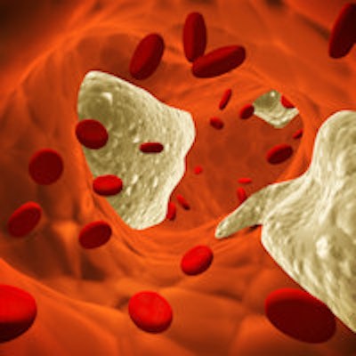
By spotting atherosclerotic plaques that aren't also present in the carotid artery, femoral ultrasound can predict the risk of cardiovascular (CV) disease in many asymptomatic patients who would be overlooked if they only received carotid ultrasound, according to research from Italy published online in Angiology.
The multicenter study included more than 300 asymptomatic adults who received ultrasound of the common femoral artery (CFA) and the common carotid artery (CCA). The team from Sapienza University of Rome found that nearly 40% of the patients with plaques had them only in their femoral artery. Many of these plaques were also lipidic, which can be treated promptly with statins, said first author Dr. Pierleone Lucatelli.
What's more, the mean intima-media thickness (IMT) measurement was higher on femoral ultrasound than on carotid ultrasound, reflecting an increased risk for stroke and CV disease.
"If confirmed by future studies, these results suggest the potential use of CFA-IMT assessment as an early biomarker of atherosclerosis and the introduction of femoral artery ultrasound in the screening protocols for cardiovascular risk assessment," wrote Lucatelli and colleagues.
CV risk assessment
Atherosclerotic lesions in the carotid artery are considered to be an indicator of generalized atherosclerosis, while carotid IMT is also an established independent predictor of stroke and CV disease, according to the authors. Because femoral ultrasound has been shown in other studies to correlate with subclinical atherosclerosis and to be an independent predictor of cardiovascular events, interest is growing in the use of the modality as an adjunctive method for stratifying cardiovascular risk (Angiology, May 27, 2016).
The group therefore sought to compare mean IMT measurements and the prevalence of atherosclerotic plaque in both the carotid and femoral arteries. The initial idea for the research project stemmed from a prior effort to investigate the heritability of carotid plaque in twins. While the multicenter study was designed to provide longitudinal follow-up for that initiative, the researchers also elected to incorporate femoral ultrasound to expand their knowledge of cardiovascular risk, Lucatelli told AuntMinnie.com.
The team invited 322 healthy participants -- 193 women and 129 men with an average age of 52.1 years -- from the Italian Twin Registry to enroll in the study at one of four different centers. All subjects received B-mode ultrasound of the supra-aortic vessels and the femoral district using a 12-MHz linear probe on a MyLab60 (Esaote), MyLab70 (Esaote), S8 (SonoScape), or Aplio XG (Toshiba Medical Systems) ultrasound scanner.
Sonographers recorded at least three IMT measurements in each segment and calculated an average IMT measurement from these results. For the purposes of the study, the researchers defined atherosclerotic plaque as an endoluminal protrusion of at least 1.5 mm or a greater than 50% focal thickening of the IMT relative to the adjacent wall segment.
Higher IMT levels
Overall, the femoral ultrasound studies found a higher level of IMT than the carotid ultrasound exams:
- Mean IMT for common femoral artery: 0.73 mm
- Mean IMT for common carotid artery: 0.70 mm
While modest, the difference was statistically significant (p = 0.0016). Given that more than one-third of plaques found in the study were detected only in the CFA, "these results suggest the hypothesis that early intima-media thickening at the common femoral site could represent a prompt marker of subclinical atherosclerosis," the authors wrote.
Overall, the prevalence of atherosclerotic plaque was 40.7% in the CFA and 30.4% in the CCA. The difference was statistically significant (p = 0.0003). While the presence of atherosclerotic plaque rose significantly in both the CFA and CCA with age, the femoral segment had a higher prevalence of plaque across all age categories, the group found.
Isolated femoral plaques
The ultrasound studies found no plaques in half of the study population. Of those who had at least one plaque, 46% had plaque in both the carotid and femoral arteries. However, 38% had plaque in only the femoral artery, while 17% had plaque only in the carotid artery.
In terms of reliably assessing cardiovascular risk in patients, "the high prevalence of isolated femoral plaque suggests that, in a certain percentage of cases, a subclinical atherosclerotic condition can be misdiagnosed in patients evaluated for carotid disease only," the authors wrote.
In addition, femoral ultrasound found more plaque of the types more amenable to statin therapy; 39% of these fibrolipid or mixed-composition plaques were found in the CFA, while 22% were found in the CCA, according to the researchers.
After comparing the performance of carotid and femoral ultrasound based on atherosclerotic risk factors, the researchers noted a number of statistically significant results.
"These findings suggest that a combined [ultrasound] examination of the two districts can improve the detection of lesions in participants with or without hypercholesterolemia, smoke habit, overweight, or higher [mean arterial pressure] values," the authors wrote.
Based on the research, the University of Rome now incorporates femoral artery ultrasound as part of its screening protocol for assessing cardiovascular risk, Lucatelli said. In future work exploring this topic, the institution would like to perform a genetics investigation if it can receive grant or investment funding, he said.