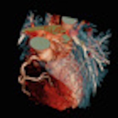
Different heart rates and rhythms have a big impact on radiation dose levels in 64-detector-row coronary CT angiography (CCTA), German researchers have found in a new study published on 22 October in the European Journal of Radiology.
Predictably, the higher doses are also related to significantly longer scan times. And unfortunately, complicated patient scenarios involving phenomena that boost the dose excessively are more common than the scanner manufacturers might wish, according to Dr. Christian Luecke, Dr. Hans Nagle, and colleagues from the University of Leipsic in Leipsic, Germany, and Wissenschaft & Technik für die Radiologie in Buccholz.
"Following the manufacturer's recommendation, prospectively gated cardiac CT should be applied in patients with sinus rhythm and low heart rate," they wrote. "In our experience, many patients do not meet these conditions, as coronary artery pathologies often are associated with higher heart rates or arrhythmias."
Prospectively gated CCTA, also known as a step-and-shoot protocol, is performed with only a slight overlap of cardiac anatomy in generally four scan steps given a detector width of 4 cm, the authors explained. In patients with heart rates below 75 beats per min (bpm), every second QRS wave complex is skipped in order to reduce the radiation dose.
Most previous studies have excluded patients with arrhythmias or high heart rates, and as a result, "the impact of ECG changes on the applied radiation exposure and scanner performance in pCT is widely unknown," they noted. The phantom study aimed to evaluate the influence of different heart rates and arrhythmias on scanner performance, image acquisition, and radiation exposure for prospectively ECG-triggered CT.
The authors of this study chose a scan range of 187.2, corresponding to a series of six scan steps in order to maximize the possibility of capturing the simulated ECG abnormalities. An ECG simulator (EKG Phantom 320, Müller & Sebastiani Elektronik, Munich, Germany) was used to generate a wide array of different heart rhythms and arrhythmias. The variants included sinus rhythm with different heart rates (45, 60, 75, 90, and 120 bpm), supraventricular extrasystoles (SVES), sinus arrhythmia, atrial flutter, atrial fibrillation, two AV blocks, intermittent asystole, mono- and polytope ventricular extrasystoles (VES), ventricular compensatory rhythm, bigeminy, R-on-T phenomenon, couplets, ventricular salvo, ST elevations and ST depression, bifocal pacemaker, demand pacemaker, pacemaker dysfunction, and technical artifacts, as well as a technical hum at 50 Hz.
Analysis of the image acquisition process was performed on a 64-detector-row scanner (Brilliance, Philips Healthcare) using a protocol that included 120 kV, 192 effective mAs, pitch of 0.78, 0.625-mm collimation, and table feed of 31.2 mm for a scan length of 187.2 mm. For simulated heart rates below 80 bpm, the researchers chose 75% of the R-R-interval inasmuch as it usually produces the fewest artifacts. For simulated heart rates above 80 bpm, systole at 45% of the R-R interval was used for the same reason. Exposure time of 0.4 seconds corresponded to about two-thirds of the data used for image acquisition and one-third for "padding."
In all, 109 scan series were performed under simulation of 26 different ECG abnormalities, including 83 repeated scan series. Fifteen were performed at 45% of the R-R interval and the rest at 75%.
| Click here to view a table of the partial results of the effect of heart rate and rhythm on dose and scan length. |
The results showed that radiation exposures can increase significantly when the heart deviates from sinus rhythm, Leucke and colleagues stated.
ECG alterations leading to significant (p < 0.05) increases in dose-length product (DLP) included the following:
- Bifocal pacemaker (61%)
- Pacemaker dysfunction (22%)
- SVES (20%)
- Ventricular salvo (20%),
- Atrial fibrillation (14%).
Significantly (p < 0.05) prolonged scan times (> 8 sec increase) could be seen in the following:
- Bifocal pacemakers (12.8 sec),
- Pacemaker dysfunction (10.7 sec)
- Atrial fibrillation (10.3 sec)
- Sinus arrhythmia (9.3 sec).
The results in each scenario can be used as a reference for deciding for or against a prospectively triggered scan in the clinical environment, Leucke and colleagues noted.
"The classification of scanner response to different ECT alterations allows estimating a potential increase in radiation exposure and an impairment of the image quality due to faulty ECG triggering, motion artifacts, and a prolonged scan time," they wrote. "The latter can result in an inadequate bolus timing and low cardiac contrast; in addition, breathing artifacts are more likely to occur."
For low heart rates, the contrast injection protocol should be adjusted, they stated. Any ECG alterations resulting in a type III scanner response including AV-block II, ventricular compensatory rhythm, bigeminy, and pacemaker dysfunction are not suitable for cardiac CT, while type IVa responses are technically suitable, but may result in a higher radiation dose.
As limitations, the authors cited the lack of a human cohort, but stated that the results should be applicable to use in a patient population. The study did not include image-quality analysis.
Changes in heart rate and rhythm "may have a significant impact on radiation exposure" and scanning time, the authors concluded. The results can serve as a guide to the feasibility of cardiac CT and also needed changes in contrast and scanning protocols, and "have an important implication for indication and informed consent," Leucke and colleagues wrote.