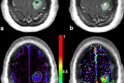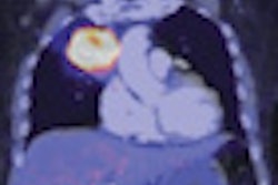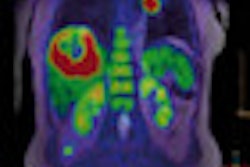
Two admittedly small studies -- one from Germany and one from Switzerland -- presented at last week's European Association of Nuclear Medicine (EANM) annual congress have underlined the emerging clinical potential of PET/MRI in both the head and neck region, and the brain.
PET/MRI of head and neck cancer is feasible with a whole-body PET/MRI system, and allows comprehensive tumor staging, noted Dr. Bettina Beuthien-Baumann, of the Klinik und Poliklinik fuer Nuklearmedizin, Universitaet Carl Gustav Carus, Dresden, Germany.
Her sample comprised 20 patients (16 men, four women, mean age 64) with histologically proven head and neck cancer. A PET scan of the body trunk was performed to rule out distant metastases, using an ECAT EXACT HR+ machine from Siemens Healthcare. A PET/MRI investigation of the head and neck region was carried out on an Ingenuity device from Philips Healthcare that incorporated a 3-tesla MR unit.
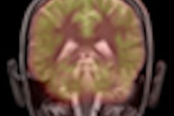
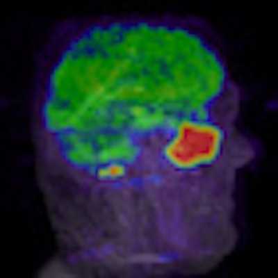
 B-cell lymphoma of the left sinus with no metastatic disease. PET/MR images are shown in all three imaging planes for a clear picture of pathology in a very critical nasal cavity area. Ingenuity TF PET/MR system was used. All images and chart courtesy of Philips Healthcare.
Of all 20 tumor locations, 17 tumors were detected with PET and MRI. Two tumors would have been missed with FDG-PET, and three tumors would have been missed by MRI. In five patients, additional lymph nodes that were suspicious for metastases were detected by FDG-PET. PET tumor detectability in the head and neck region increased on the late imaging on the PET-MR system, which was due to the increased tumor contrast at a later imaging time point and the higher resolution of the new device. Although a direct comparison of standard uptake values (SUV) of a different PET scanner is not valid, in parallel to the increased qualitative detectability, at late timepoints an increase of SUVmax values of the tumors was seen, according to the Dresden team.
"PET/MRI systems combine the unique metabolic imaging capabilities of PET and the excellent tissue contrast of MRI," Beuthien-Baumann stated. "It is anticipated that in head and neck cancer, the sensitivity and specificity of the detection of lymph node metastases might be ameliorated with such a combined system."
In the second EANM study, researchers from Geneva University Hospital in Switzerland, tested a new whole-body hybrid PET/MR system to evaluate the performance and clinical applicability of combined imaging protocols for brain studies.
Acquiring both PET and MRI in a single session on a hybrid device can minimize patient discomfort while maximizing clinical information and optimizing registration of both modalities, according to lead author Dr. Valentina Garibotto, from the department of medical imaging. Simplification of imaging protocols allowed keeping the total duration of the examination within a tolerable range (less than two hours), which has an important impact on image quality. In comparison to PET/CT, the lower effective dose is particularly beneficial for children, she pointed out.
 Dr. Osman Ratib, PhD, professor and chair of radiology at University Hospital of Geneva, prepares a patient for a PET/MRI examination.
The latest PET/MR systems use different solutions to overcome the interference of the magnetic field on the electronic detection of the PET signal, both for brain dedicated as well as whole-body imaging, using a sequential or a synchronous acquisition scheme.
"One of the major challenges in this new technology is optimization of acquisition protocols in order to obtain diagnostic quality images in a clinically acceptable time frame for both modalities, including specific MR sequences, such as spectroscopy, diffusion tensor imaging, perfusion imaging, and functional MRI," she noted.
A total of 17 patients (10 men, seven women, mean age 38) were examined using a Philips Ingenuity TF PET/MR system. The device consists of two separate machines linked through a single patient table, allowing sequential imaging. Standard imaging protocols of both modalities were combined.
In all cases, diagnostic quality images were obtained for both modalities in a single session. A total of 13 subjects had positive findings, which were confirmed by clinical follow-up in seven cases. One false positive PET uptake was due to radionecrosis. The remaining five positive subjects showed metabolic changes, the interpretation of which is currently under evaluation with clinical follow-up (e.g., global cortical hypometabolism and basal ganglia hypermetabolism in a case of obsessive compulsive disorder). Of the four negative studies, no false negatives were identified with clinical follow-up.
"We also tested a specific MR-compatible EEG recording system in order to obtain simultaneous EEG monitoring of epileptic patients during functional MRI and PET acquisition. Images were interpreted by multidisciplinary teams of nuclear physicians, radiologists, and neurologists," Garibotto commented.
','dvPres', 'clsTopBtn', 'true' );">

Click to view chart.