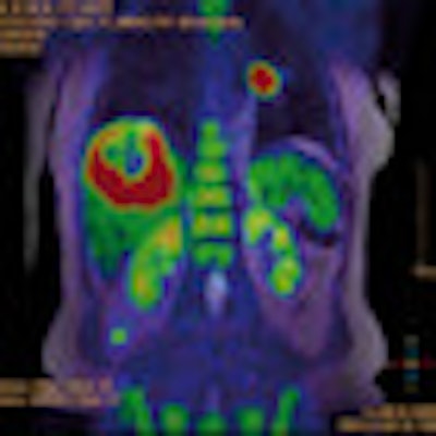
CHICAGO - PET/MRI can outperform PET/CT for lesion conspicuity and characterization in the abdomen, particularly in the liver, according to a study presented by Swiss researchers on Wednesday at the RSNA 2011 meeting.
A total of 19 oncology patients with a mean age of 55 years were enrolled in the study, said lead study author Dr. David Crook, from University Hospital Zurich. Inclusion criteria were a clinically indicated FDG-PET/CT examination for staging and restaging of cancer, a lack of contraindications for PET/MRI, and the presence of a PET-positive lesion in the abdomen.
In the first part of imaging protocol, all 19 patients received a time-of-flight (TOF) PET/CT scan (Discovery PET/CT 690, GE Healthcare) with low-dose CT for attenuation correction. No contrast agent was administered unless requested by the referring physician.
Patients then completed a 3-tesla MRI exam (Discovery MR750, GE Healthcare), with researchers obtaining a T1-weighted 3D gradient recalled echo (GRE) image of the abdomen. Once again, no contrast agent was given unless requested.
A connecting patient transfer table allowed the use of a 32-channel torso coil after PET/CT to avoid coil-induced CT and PET attenuation correction artifacts and to maintain a stable patient position.
Two double-board-certified radiologists and nuclear physicians analyzed PET/MRI and PET/CT data independently, grading PET-positive lesions on a three-point scale for lesion conspicuity in CT and MRI.
A total of 27 PET-positive lesions were evaluated on PET/CT and PET/MRI; the majority of lesions were found in the liver (12), followed by bone (four), the stomach/bowel region (three), pleura (two), lung (two), fatty tissue (two), pancreas (one), and epidural space (one).
Lesions were characterized with moderate and good interobserver agreement on lesion conspicuity in CT and MR, respectively, Crook noted. In an overall lesion-based comparison, no significant difference was found, although a tendency for MRI to outperform CT was apparent (p = 0.11 and 0.08, respectively).
"In comparing liver lesions, MR significantly outperformed CT," Crook added. "However, in extra fatty [tissue] findings, there was no significant difference found in this patient population."
The preliminary data indicate that PET/MRI "tends to outperform PET/CT in abdominal lesion characterization and conspicuity when using unenhanced CT and MRI, although this difference falls short of statistical significance," Crook and colleagues concluded. The difference, however, is significant in the liver.
Crook recommended that further research be done with additional MR sequences to fully evaluate the potential of PET/MRI compared to PET/CT.
| Crook receives R&D funding from GE Healthcare. |
Editor's note: The image on the home page is courtesy of the University of Geneva.


![Examples of ultrasound findings and techniques. (A) Images in a 39-year-old male patient with a mass in the left thigh. The mass is heterogeneous on the B-mode US image (compared with the patient in D) and showed increased microvascularity (superb microvascular imaging [SMI]) and shear-wave elastography (SWE) values. Undifferentiated pleomorphic sarcoma was diagnosed at biopsy (with pleomorphic rhabdomyosarcoma in surgical specimen). (B) Images in an 18-year-old male patient with a mass in the left leg. The mass is hypoechoic on the B-mode image, with no other findings suggestive of malignancy. The lesion is in contact with the cortex of the tibia, which is slightly irregular. CT revealed a doubtful anteromedial tibial erosion. The microvascular study demonstrated high vascularization, suggestive of malignancy. Periosteal Ewing sarcoma was diagnosed with both histologic and immunohistochemical confirmation. (C) Images in a 69-year-old female patient with a lump growing on the outside of the left leg. Multiple SWE examinations were performed (please note the high values obtained in the measurements, whereas the color map highlights the stiffness relative to adjacent tissues). SMI showed areas of increased vascularization to target for sampling. Undifferentiated spindle cell sarcoma was diagnosed at biopsy, with residual leiomyosarcoma in the surgical specimen after neoadjuvant therapy. (D) Images in a 56-year-old female patient with a mass in the right thigh. The mass is heterogeneous at both B-mode ultrasound (similar to patient A) and MRI (coronal T2-weighted spectral attenuated inversion recovery [SPAIR]; T1-weighted pre-contrast and postcontrast imaging), which even shows uptake after the administration of paramagnetic contrast material, which is traditionally suggestive of malignancy. Low values at SMI and elastography are suggestive of benignity. Spindle cell lipoma was diagnosed at biopsy, with atypical spindle cell lipomatous tumor in the surgical specimen.](https://img.auntminnieeurope.com/mindful/smg/workspaces/default/uploads/2026/08/images-radiol250278fig2.APCFLSvX6p.jpg?auto=format%2Ccompress&fit=crop&h=100&q=70&w=100)


![Examples of ultrasound findings and techniques. (A) Images in a 39-year-old male patient with a mass in the left thigh. The mass is heterogeneous on the B-mode US image (compared with the patient in D) and showed increased microvascularity (superb microvascular imaging [SMI]) and shear-wave elastography (SWE) values. Undifferentiated pleomorphic sarcoma was diagnosed at biopsy (with pleomorphic rhabdomyosarcoma in surgical specimen). (B) Images in an 18-year-old male patient with a mass in the left leg. The mass is hypoechoic on the B-mode image, with no other findings suggestive of malignancy. The lesion is in contact with the cortex of the tibia, which is slightly irregular. CT revealed a doubtful anteromedial tibial erosion. The microvascular study demonstrated high vascularization, suggestive of malignancy. Periosteal Ewing sarcoma was diagnosed with both histologic and immunohistochemical confirmation. (C) Images in a 69-year-old female patient with a lump growing on the outside of the left leg. Multiple SWE examinations were performed (please note the high values obtained in the measurements, whereas the color map highlights the stiffness relative to adjacent tissues). SMI showed areas of increased vascularization to target for sampling. Undifferentiated spindle cell sarcoma was diagnosed at biopsy, with residual leiomyosarcoma in the surgical specimen after neoadjuvant therapy. (D) Images in a 56-year-old female patient with a mass in the right thigh. The mass is heterogeneous at both B-mode ultrasound (similar to patient A) and MRI (coronal T2-weighted spectral attenuated inversion recovery [SPAIR]; T1-weighted pre-contrast and postcontrast imaging), which even shows uptake after the administration of paramagnetic contrast material, which is traditionally suggestive of malignancy. Low values at SMI and elastography are suggestive of benignity. Spindle cell lipoma was diagnosed at biopsy, with atypical spindle cell lipomatous tumor in the surgical specimen.](https://img.auntminnieeurope.com/mindful/smg/workspaces/default/uploads/2026/08/images-radiol250278fig2.APCFLSvX6p.jpg?auto=format%2Ccompress&fit=crop&h=112&q=70&w=112)