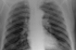
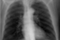
Mobile chest digital radiography (DR) provides superior image quality to computed radiography (CR) at the bedside, Dutch researchers found in a study published this month in Clinical Radiology. In addition, DR studies produce 50% less radiation dose without the loss of diagnostic information.
CR is widely available and generally accepted for both upright and bedside chest radiography, but recently DR has become increasingly used for upright chest radiography. DR offers improved detective quantum efficiency (DQE) that can be used to reduce radiation dose while maintaining image quality, or to increase the signal-to-noise ratio and image quality.
However, mobile DR units have lower quantum efficiency, thus the radiation dose reduction may not be as significant as that reported for upright DR units. So far publications on mobile DR chest radiography have been limited to pediatric applications. Therefore, the aim of the current study was to compare mobile DR units with mobile CR units for adult bedside chest radiography with respect to image quality, dose reduction, and the effect on diagnostic performance.
Dr. Diederick De Boo from the radiology department at the Academic Medical Center in Amsterdam and colleagues compared three groups of age-, weight-, and disease-matched intensive care unit patients. Images for the 114 patients were obtained with either CR (Agfa HealthCare), DR (Mobilett XP Digital, Siemens Healthcare), or DR with a 50% dose reduction. Fifty chest x-rays were obtained for each acquisition technique (Clin Radiol, 12 May 2011).
Four readers scored delineation of anatomic structures and devices used for patient monitoring, overall image quality, and disease. In 12 patients, pairs of follow-up CR and DR images were available, and in 15 patients pairs of CR and DR with a 50% dose reduction images were available. In the pairs, image quality was compared side by side.
The researchers found delineation of anatomy in the mediastinum was scored better with DR or DR at 50% than with CR. Devices used for patient monitoring were seen best with DR, as well as with DR at 50%. In the side-by-side comparison, the overall image quality of DR and DR at 50% was rated better than CR in 96% and 87% of cases. Interobserver agreement for pathology assessment was fair for CR and reduced-dose DR, and moderate for DR.
The study shows despite the dose reduction in DR, the device still outperformed CR for some imaging aspects.
"In order to be able to place these dose considerations into perspective, it is important to state not only relative but also absolute dose levels: Reduction of a relatively high 'standard dose' by a factor of two seems to be less of an achievement compared with operating on a generally lower dose level," the researchers wrote.
In the current study, the standard dose amounted to a mean of 0.18 mGy entrance skin dose for CR and DR and 0.09 mGy entrance skin dose for DR at 50%, which are comparable with other studies of upright chest radiography.
While the levels are lower compared with conventional film-screen x-ray, dose reduction remains a relevant issue, as patients often require multiple follow-up studies, the researchers noted.
The researchers also note image quality requirements are different in bedside chest radiography compared with routine upright chest radiography. Contrast resolution has to be high to display the multiple types of catheter materials, whereas requirements for spatial resolution are generally low. Even so, DR was found to be superior to CR in the current study.
"These results confirm previous experiences that DR either allows for improved image quality and potentially improved diagnostic performance if obtained with the same dose as that used for CR, or alternatively allows for dose reduction on a slightly lower but unaltered diagnostic level comparable to CR," the researchers stated in their study.
However, the study suffers from some limitations: First, the catheter fragments were relatively short and superimposed over the area of the upper abdomen and not within the mediastinum. Therefore, position and length of the catheters lacked clinical characteristics, but this technique allowed the visualization of a range of monitoring devices to be tested under controlled conditions.
Second, image pairs of the same patient obtained with two acquisition techniques were available in only a subgroup of the study patients.
Third, acquisition parameters were set by the technicians according to the patient's weight and the technician's visual assessment of the patient's constitution and condition.
"Although it cannot be proved that every single DR image was acquired with exactly 50% less dose, on average DR images had been obtained either with the 'standard' or with a dose lowered by 50%," the researchers wrote.
Despite the limitations, the researchers still conclude DR outperformed CR at the bedside.


