Photoacoustic technology, the use of light-induced sound, is showing promise in imaging of breast cancer. It may be able to distinguish malignant tissue from normal tissue by providing high-contrast images of tumors, Dutch researchers said.
Preliminary results of the first phase of clinical testing of a novel imaging device, the Twente Photoacoustic Mammoscope (PAM), are described in the 21 May issue of Optics Express. The images generated from 12 patients with diagnosed breast cancer provide proof-of-concept support that the technology may be viable (Optics Express, June 2012, Vol. 20:11, pp. 11582-11597).
The research uses light-induced sound produced by the device, rather than ionizing radiation, to detect and visualize breast tumors. It was undertaken by researchers from the University of Twente and the Medisch Spectrum Twente Hospital in Oldenzaal, the Netherlands.
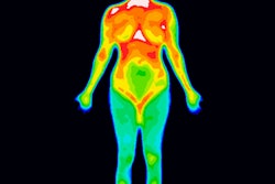
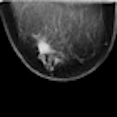
 Mixed infiltrating lobular and ductal carcinoma in right breast of 57-year-old patient. The craniocaudal x-ray mammogram (left) shows an architectural distortion of about 22 mm in the lateral part of the right breast. Ultrasound (middle) shows the presence of an smooth-edged hypoechoic lesion with a hyperechoic border at the expected location. Photoacoustic mammography (right) shows a confined high-contrast abnormality with a contrast in excess of 5 mm and a maximum diameter of 14 mm at the expected lesion depth. Here, a transversal cross-section through this abnormality is visualized. All images courtesy of Michelle Heijblom, University of Twente.
Photoacoustics, a hybrid optical and acoustical imaging technique, builds on the established technology of using red and infrared light to image tissue and detect tumors. Optical mammography reveals malignancies because blood hemoglobin readily absorbs the longer, redder wavelengths of light. These reveal a clear contrast between blood-vessel dense tumor areas and normal vessel environments.
However, because it is difficult to target a specific area to be imaged using this approach, the PAM combines a light-based system's ability to distinguish between benign and malignant tissue with ultrasound to achieve superior targeting ability.
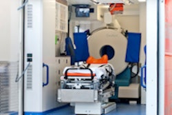
Lead author Michelle Heijblom, a doctoral student at the University of Twente, and colleagues, explained that the device is built into a hospital bed, where the patient lies prone. Laser light at a wavelength of 1,064 nanometers scans the breast. Because there is an increased absorption of the light in malignant tissue, the temperature increases slightly. With a rise in temperature, thermal expansion creates a pressure wave, which is detected by an ultrasound detector placed on one side of the breast.
 The Twente Photoacustic Mammoscope.
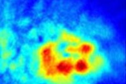
The resulting photoacoustic signals are then processed by the PAM system and reconstructed into images. These images reveal abnormal areas of high intensity that are tumor tissue as compared to areas of low intensity that are benign tissue.
By comparing the photoacoustic data with conventional diagnostic mammography images, ultrasound imaging, breast MRI, and tissue examinations, the researchers showed that malignancies produced a distinct photoacoustic signal which has the potential to be clinically useful in making a diagnosis of breast cancer. The research team also observed that the photoacoustic contrast of the malignant tissue was higher than the contrast provided by conventional mammography images.
Heijblom and colleagues said they think some technical improvements of the PAM system need to be made. "Our next step is to make those improvements and then use it to evaluate less obvious potential tumors, benign lesions, and normal breasts," she noted.