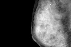
Ultrasound and x-ray mammography have several limitations for the detection and monitoring of breast cancer, including high false-positive rates and exposure to ionizing radiation, and this has motivated research into alternatives such as photoacoustic (PA) imaging.
In a step toward identifying the clinical utility of PA imaging, researchers in the Netherlands have detected breast lesions in PA images at locations predicted by conventional imaging. Correctly identified in 28 out of 29 patients, the lesions exhibited distinctive patterns that the authors hypothesize result from the abnormally dense microvasculature associated with tumors (Scientific Reports, 10 July 2015, Vol. 5).
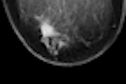
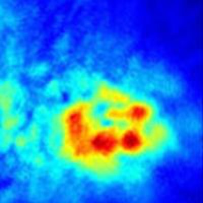
 Contrast-enhanced MR images (left) are compared with PA images (right) in three lesions. The dashed box denotes the area scanned using the PAM system. Credit: Scientific Reports.
"Our study shows that PA imaging has potential in visualizing breast cancer," said first author Michelle Heijblom, PhD, of the MIRA Institute at the University of Twente in Enschede.
"Investigations of the appearance of breast lesions should be followed up in a large cohort," added senior author Srirang Manohar, PhD. "This could contribute to the development of PA image descriptors that could eventually act as diagnostic criteria for the technique." If achieved, the approach would also be an alternative to contrast-enhanced MRI that, while sensitive, is expensive, requires injection of a contrast agent, and lacks specificity.
Manohar and colleagues carried out the study using an upgraded version of the Twente Photoacoustic Mammoscope (PAM), an in-house system first developed in 2007. The system comprises a pulsed Nd:YAG laser and a flat 588-element 2D ultrasound detector, enabling partial scans of the breast.
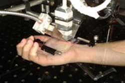
 First author Michelle Heijblom and senior author Srirang Manohar. Image credit: R Molenaar.
Patients are scanned prone with their breast positioned pendant through an aperture and compressed cranio-caudally between the detector and a glass window. The laser is fired through the window and the beam is expanded to a diameter of around 70 mm to illuminate the breast. The resulting ultrasound is measured by the detector and an acoustic back projection algorithm reconstructs 3D PA images.
The researchers scanned patients with suspicious lesions detected by conventional imaging. The cohort revealed three different PA intensity patterns. One was mass-like with a high-intensity core. A second was that of a full or partial ring of high intensity with a region of low intensity inside. A third "nonmass" pattern constituted a scattering of high-intensity foci over an extended region. Though further investigation is required to confirm the finding, the patterns are thought to indicate tumor vasculature, as at the 1,064 nm laser wavelength used, hemoglobin and oxyhemoglobin are the dominant molecular absorbers.
 PA images showing a ring-like lesion (middle) are compared with conventional clinical images (top) and H&E and CD-31 stained tissue sections (bottom) showing tumor cell and vascular endothelial cells. Credit: Scientific Reports.
In eight small-to-medium size lesions, MR images, which show the transit of contrast agent through the vasculature, were consistent with this: seven had shapes and patterns similar to the PA images, a visual inspection revealed. In all cases where MR images where available, lesions were also in the same anatomical locations.
Qualitative comparisons of the PA images over the whole cohort with H&E stained tissue sections demonstrating tumor cell distribution also revealed similarities in lesion shape, size, and pattern. In six cases, a further histological analysis of tissue stained to visualize microvasculature also demonstrated similarities to the PA images.
Based on their findings, the PA technique has potential in clinical breast imaging, though more research is needed, Manohar told medicalphysicsweb. "In an optimistic scenario, our method can be used in niche areas in approximately five to 10 years," he said. "But it could take longer before the method can be used regularly for screening and diagnosis."
 The breast is positioned pendant through an aperture and compressed cranio-caudally between the ultrasound detector and a glass window. The laser is fired through the window and expanded to a diameter of around 70 mm to illuminate the breast. Credit: Scientific Reports.
In ongoing work, the researchers are developing a new version of their system, PAM 2, that will use two wavelengths of light for improved tissue characterization and more advanced electronics to perform quicker scans, of the whole breast. Expected to be ready by the end of this year, PAM 2 will be used in the group's first blinded clinical PA study.
© IOP Publishing Limited. Republished with permission from medicalphysicsweb, a community website covering fundamental research and emerging technologies in medical imaging and radiation therapy.