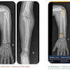
Using a photon-counting CT system for coronary CT angiography (CCTA) improves image quality and diagnostic confidence among readers compared with conventional CT, according to a study published February 15 in Radiology.
Photon-counting CT offers a number of benefits compared with traditional CT for CCTA for coronary artery disease (CAD), wrote a team led by Dr. Salim Si-Mohamed, PhD, of the University Lyon in Villeurbanne, France.
"Spatial resolution, soft-tissue contrast, and dose-efficient capabilities of photon-counting CT potentially allow a better quality and diagnostic confidence of coronary CT angiography in comparison to conventional CT," the team noted.
CCTA is the go-to exam for assessing cardiovascular disease and has a high negative predictive value for CAD, Si-Mohamed and colleagues wrote. But its spatial resolution and soft-tissue contrast limitations can affect diagnostic capability when it comes to small arteries and high-contrast (stents, calcifications) and low-contrast (noncalcified plaque) imaging. It also imparts a relatively high dose of radiation.
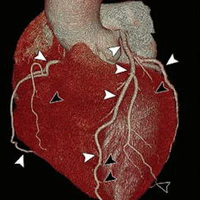
 Images from (A-E) coronary photon-counting CT and (F-J) energy-integrating detector dual-layer CT angiography in a 44-year-old woman. Volume-rendered photon-counting CT image (A) and volume-rendered energy-integrating detector dual-layer CT image (F) depict proximal coronary arteries (white arrowheads), but there is clear improvement in the depiction of distal coronary arteries (black arrowheads in A) with volume rendering and photon-counting CT in comparison to energy-integrating detector dual-layer CT. On axial images, pectinate muscle (B, G), aortic cusp commissure (C, H), noncoronary cusp (D, I), and papillary muscle (E, J) were better depicted on photon-counting CT images (B-E) than on energy-integrating detector dual-layer CT images (G-J) (white arrowheads). Black arrowheads indicate distal coronary arteries not depicted on energy-integrating detector dual-layer CT images. Images and caption courtesy of the RSNA.
Photon-counting CT addresses these concerns by using "energy-resolving" (i.e., photon-counting) detectors that separately track the energy of each photon, according to the authors. The U.S. Food and Drug Administration (FDA) cleared the first device in September 2021 (Naeotom Alpha, Siemens Healthineers), and since then, there has been an increasing number of investigations into clinical uses of the technology.
But photon-counting CT hasn't yet been tested for use for CCTA. Si-Mohamed and colleagues performed what they said is the first human study to do so.
The research included 14 individuals with coronary artery disease who underwent electrocardiographically gated, contrast-enhanced CCTA on both a clinical prototype photon-counting CT device and a typical energy-integrating detector dual-layer CT system. Three cardiac radiologists read the exams and evaluated them for image quality, diagnostic confidence, and diagnostic quality of any features such as calcifications, stents, and noncalcified plaques using a five-point scale (1 = insufficient, 5 = excellent).
The group found that overall scores for image quality and diagnostic confidence were higher on the photon-counting CT images compared with images produced by a conventional CT system (5 vs. 4). Expressed in percentage of quality score improvement, the group reported the following:
| Percentage quality score improvement with photon-counting CT compared with conventional CT for CCTA | |
| Measure | Percentage improvement |
| Overall image quality | 57% |
| Diagnostic confidence | 55% |
| Diagnostic quality | |
| Calcifications | 100% |
| Stents | 92% |
| Noncalcified plaque | 45% |
| Proximal lumen | 48% |
| Distal lumen | 51% |
| Coronary wall | 69% |
The study findings highlight the promise of photon-counting CT both for cardiac applications and for other imaging, according to a commentary accompanying the study by Dr. Veit Sandfort of Stanford University and Dr. David Bluemke, PhD, of the University of Wisconsin in Madison and editor of Radiology.
"This technology will take time to spread, but the inventiveness of the imaging community is profound," Sandfort and Bluemke wrote. "In the past 30 years, we have experienced helical CT, wide-detector CT, and spectral CT. Each time, it seemed CT may have peaked. Once again, CT has reinvented itself with photon-counting detectors."