Dear AuntMinnieEurope Member,
Understandably, Dutch radiologists still feel angry about a senior politician's recent statement that they're going to be replaced by artificial intelligence (AI) technology.
Finance Minister Wopke Hoekstra referred to radiologists as "mensen," or humans, instead of physicians or medical specialists, and in the next breath he mentioned that similar changes were happening to supermarket checkout staff. Considering Hoekstra's wife, Liselot Hoornweg, has worked as a general practitioner for nearly eight years, his comments seem remarkably ill-informed and insensitive.
Dr. Merel Huisman, PhD, a fourth-year radiology resident, has entered the debate and presented her views about the profession's future. As an epidemiologist and a board member of the European Society of Medical Imaging Informatics, she is worth listening to. Find out more in the Artificial Intelligence Community.
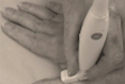
The Alpine Skiing World Cup begins in Austria this weekend, so now is an ideal time to brush up on your diagnostic knowledge of skier's thumb. These acute injuries are relatively common and were addressed recently at the virtual ultrasound workshop hosted by the European Society of Musculoskeletal Radiology.
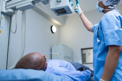
Breast cancer patients with high levels of coronary artery calcium at the outset of treatment have a higher risk of later hospitalization or death from cardiovascular disease. That's the key finding from a new study presented at the 2020 European Breast Cancer Conference. Visit the Women's Imaging Community.
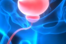
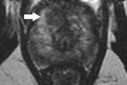
MRI has become a valuable tool for detecting, localizing, and staging prostate cancer, but the introduction of prebiopsy scans in suspected cases of malignancy has exerted a huge strain on healthcare systems, and the pressure is on imaging departments to report exams quickly. Radiologists in the north of Scotland have embarked on a rigorous campaign to speed up the booking and reporting process. Don't miss our news report in the MRI Community.


![Overview of the study design. (A) The fully automated deep learning framework was developed to estimate body composition (BC) (defined as subcutaneous adipose tissue [SAT] in liters; visceral adipose tissue [VAT] in liters; skeletal muscle [SM] in liters; SM fat fraction [SMFF] as a percentage; and intramuscular adipose tissue [IMAT] in deciliters) from MRI. The fully automated framework comprised one model (model 1) to quantify different BC measures (SAT, VAT, SM, SMFF, and IMAT) as three-dimensional (3D) measures from whole-body MRI scans. The second model (model 2) was trained to identify standardized anatomic landmarks along the craniocaudal body axis (z coordinate field), which allowed for subdividing the whole-body measures into different subregions typically examined on clinical routine MRI scans (chest, abdomen, and pelvis). (B) BC was quantified from whole-body MRI in over 66,000 individuals from two large population-based cohort studies, the UK Biobank (UKB) (36,317 individuals) and the German National Cohort (NAKO) (30,291 individuals). Bar graphs show age distribution by sex and cohort. BMI = body mass index. (C) After the performance assessment of the fully automated framework, the change in BC measures, distributions, and profiles across age decades were investigated. Age-, sex-, and height-adjusted body composition reference curves were calculated and made publicly available in a web-based z-score calculator (https://circ-ml.github.io).](https://img.auntminnieeurope.com/mindful/smg/workspaces/default/uploads/2026/05/body-comp.XgAjTfPj1W.jpg?auto=format%2Ccompress&fit=crop&h=100&q=70&w=100)


![Overview of the study design. (A) The fully automated deep learning framework was developed to estimate body composition (BC) (defined as subcutaneous adipose tissue [SAT] in liters; visceral adipose tissue [VAT] in liters; skeletal muscle [SM] in liters; SM fat fraction [SMFF] as a percentage; and intramuscular adipose tissue [IMAT] in deciliters) from MRI. The fully automated framework comprised one model (model 1) to quantify different BC measures (SAT, VAT, SM, SMFF, and IMAT) as three-dimensional (3D) measures from whole-body MRI scans. The second model (model 2) was trained to identify standardized anatomic landmarks along the craniocaudal body axis (z coordinate field), which allowed for subdividing the whole-body measures into different subregions typically examined on clinical routine MRI scans (chest, abdomen, and pelvis). (B) BC was quantified from whole-body MRI in over 66,000 individuals from two large population-based cohort studies, the UK Biobank (UKB) (36,317 individuals) and the German National Cohort (NAKO) (30,291 individuals). Bar graphs show age distribution by sex and cohort. BMI = body mass index. (C) After the performance assessment of the fully automated framework, the change in BC measures, distributions, and profiles across age decades were investigated. Age-, sex-, and height-adjusted body composition reference curves were calculated and made publicly available in a web-based z-score calculator (https://circ-ml.github.io).](https://img.auntminnieeurope.com/mindful/smg/workspaces/default/uploads/2026/05/body-comp.XgAjTfPj1W.jpg?auto=format%2Ccompress&fit=crop&h=112&q=70&w=112)


