

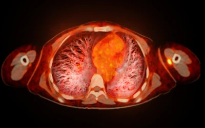
SPECT and PET imaging have become specialized disciplines for diagnosing cardiac amyloidosis, with SPECT/CT significantly outperforming traditional planar imaging by reducing false positives by up to 48%, while PET tracers like florbetaben achieve 93% diagnostic accuracy and enable better disease characterization and treatment monitoring.
- SPECT/CT superiority: Planar imaging produces false-positive results in up to 48% of cases with grade 1 findings, while SPECT eliminates this confusion between blood-pool activity and true myocardial uptake
- Tracer selection matters: PYP and HMDP are effective agents for cardiac amyloidosis imaging, while MDP produces false-negative findings and is unsuitable for this application
- PET diagnostic accuracy: The CAPRI study showed 18F-florbetaben PET achieved 90% accuracy using cardiac variables alone and 93% when combined with extracardiac variables
- Clinical practice shift: Japanese guidelines now permit tafamidis and acoramidis prescription without mandatory biopsy following a 2025 working-group report
- H/CL ratio limitations: Heart-to-contralateral lung ratios should serve as supplementary tools to visual assessment rather than stand-alone diagnostic criteria
Cardiac amyloidosis imaging has become a distinct discipline rather than simply a byproduct of routine bone scintigraphy, argued two presentations during a live webinar held on Friday by the Japanese Society of Nuclear Medicine (JSNM)/European School of Multimodality Imaging and Therapy (ESMIT).
Dr. Takashi Kudo of the Department of Radioisotope Medicine, Atomic Bomb Disease Institute, Nagasaki University in Nagasaki, Japan, urged the field to distinguish cardiac amyloid radionuclide imaging from conventional bone scintigraphy. He proposed the term “CARI,” for cardiac amyloid radionuclide imaging, and stressed the importance of SPECT.
SPECT moves beyond planar imaging
His central message was: use SPECT, and preferably SPECT/CT. Planar imaging can confuse blood-pool activity with true myocardial uptake and lead to false-positive findings. Kudo presented data showing that among patients with no uptake on SPECT, 14% appeared positive with grade 2 or 3 uptake on planar imaging. When grade 1 findings were included, the false-positive proportion rose to 48%. Planar imaging may also miss focal myocardial uptake and therefore produce false-negative results.
Kudo also cautioned against relying on the heart-to-contralateral lung, or H/CL, ratio as a stand-alone diagnostic criterion. Instead, he presented it as a supplementary tool to visual assessment, noting that activity in the contralateral lung can substantially influence the measurement.
Tracer selection was another practical focus. PYP and HMDP were identified as useful agents, whereas MDP can produce false-negative findings and was described as unsuitable for cardiac amyloidosis imaging.
Kudo also reported a change in Japanese practice following a Japanese Society of Nuclear Cardiology working-group report published in 2025. Six months after publication of the report, he said, tafamidis and acoramidis could be prescribed without mandatory biopsy.
PET adds disease characterization
Dr. Pierre-Yves Marie of the Department of Nuclear Medicine at University Hospital Nancy and Université de Lorraine in Nancy, France, then reviewed amyloid-binding PET tracers for earlier detection, disease characterization, and follow-up.
Marie compared F-18 labelled thioflavin derivatives, including florbetapir and florbetaben, with the investigational tracer 124I-evuzamitide. Both approaches showed potential for cardiac amyloidosis diagnosis, partial differentiation of AL from ATTR disease, and treatment monitoring, although their practical characteristics differ.
In the multicenter CAPRI study, early whole-body F-18 florbetaben PET achieved about 90% overall accuracy using cardiac variables alone and 93% when cardiac and extracardiac variables were combined.
Marie said two phase III studies aimed at market authorization have been completed but remain unpublished. Despite progress in PET, he concluded that biopsy-free differentiation between AL and ATTR cardiac amyloidosis will probably continue to rely on bone scintigraphy in the near term, within a broader diagnostic work-up that also excludes AL amyloidosis.