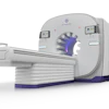
Photon-counting CT (PCCT) produces superior image quality at lower radiation doses compared to traditional CT scans in patients with obesity, according to a new study published in Radiology. The technology directly converts individual photons into electronic signals, reducing noise and improving diagnostic confidence while enabling safer, more frequent follow-up imaging.
- Superior Image Quality: PCCT demonstrated higher signal-to-noise ratio in vascular structures (aorta SNR 19.65 vs 13.22) and organs compared to energy-integrating detector CT
- Lower Radiation Dose: PCCT reduced volume CT dose index (11.99 mGy vs 17.05 mGy) and dose length product by approximately 26% compared to traditional CT
- Better Contrast and Sharpness: Both radiologists rated PCCT superior in overall image quality, contrast, and sharpness with reduced artifacts
- Improved Performance in Obesity: Signal-to-noise ratio advantages increased with higher body mass index, with stronger benefits in vessels and organs
Photon-counting CT (PCCT) could yield better image quality in patients with obesity, according to research published October 20 in Radiology.
PCCT resulted in superior quantitative and subjective image quality at a lower radiation dose compared to energy-integrating detector (EID) CT, wrote a team led by Jan Hegmanns, an MD student, from University Hospital Essen in Germany.
“These findings suggest that PCCT might play an important role in imaging of patients with obesity by enhancing diagnostic confidence, improving the detection of clinically relevant findings, and offering substantial potential for further dose optimization in future applications,” the Hegmanns team wrote.
Imaging patients with higher body mass index (BMI) can be challenging to x-ray attenuation increasing, which leads to low-quality CT images. PCCT can address this by directly converting individual photons into electronic signals, reducing noise.
However, the researchers noted limited data comparing PCCT with the established EID CT, including in settings where CT systems face technical constraints, such as imaging patients with obesity.
Hegmanns and colleagues compared radiation dose, quantitative image quality, and subjective image quality between both imaging techniques in this patient population. The study included 51 patients with obesity who underwent thoracic and/or abdominal PCCT and EID CT between 2022 and 2025. Two radiologists independently rated image quality on a six-point Likert scale.
 Representative abdominal CT images in three patients with obesity and increasing body mass index (BMI; calculated as patient weight in kilograms divided by patient height in meters squared). Contrast-enhanced axial abdominal CT images in the venous phase are shown for three patients undergoing clinically indicated routine examinations with (A–C) energy-integrating detector (EID) CT (top row) and (D–F) photon-counting CT (PCCT) (bottom row); each column represents one patient, and BMI increases from left to right. Volume CT dose index (CTDIvol) is displayed on each image to document the radiation dose of the individual examination. (A, D) Images in a 46-year-old male patient; CTDIvol is 12.43 mGy at EID CT and 10.32 mGy at PCCT, and BMI is 34.79. (B, E) Images in a 69-year-old female patient; CTDIvol is 24.24 mGy at EID CT and 16.39 mGy at PCCT, and BMI is 42.52. (C, F) Images in a 54-year-old female patient; CTDIvol is 29.44 mGy at EID CT and 18.98 mGy at PCCT, and BMI is 49.21. Window center is 50 HU, and window width is 340 HU. In each of the three patients, the PCCT images were acquired at a lower CTDIvol than the corresponding EID CT images (P < .001).RSNA
The researchers reported high signal through PCCT in vascular structures, parenchymal organs, and muscle and more negative in subcutaneous fat (all p < 0.001), with similar image noise (p = 0.09 to 0.84).
PCCT also yielded a higher signal-to-noise ratio (SNR) in vascular structures. The ascending aorta had a mean SNR of 19.65 on PCCT compared to 13.22 for EID CT (p < 0.001). And for the liver, SNR on PCCT was 5.53 compared to 4.71 on EID CT (p = 0.02). The difference in SNR increased with body mass index for vessels (r = 0.36) and parenchymal organs (r = 0.51, both p < 0.001).
Both readers rated overall image quality, contrast, and sharpness higher and artifact-related impairment lower for PCCT (all p < 0.001). However, subjective image noise was similar (p = 0.16 and 0.14).
Finally, compared to EID CT, PCCT use led to lower volume CT dose index (median, 11.99 vs. 17.05 mGy) and dose length product (median, 526.25 vs 709.32 mGy · cm; both p < 0.001).
The results shed light on the theoretical and phantom-based advantages of PCCT that could translate into measurable clinical benefits in patients with obesity, the study authors highlighted.
“The combination of meaningful dose reduction and improved SNR may support safer longitudinal imaging strategies in patient groups requiring frequent follow-up,” they wrote. “Our findings further indicate that additional dose reduction may be feasible without compromising diagnostic integrity.”
Read the full study here.