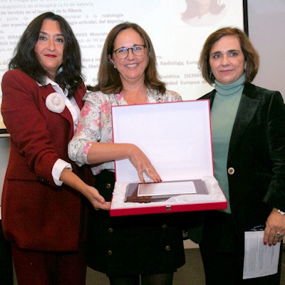
The Spanish Society of Medical Radiology (SERAM) and the Spanish Radiology Foundation (FER) have handed out the Radiology 2022 awards.
The awards recognize the valuable contributions of professionals to industry as well as other outstanding achievements and contributions, noted SERAM in a press release. The event formed part of Spanish radiology's celebrations to mark the International Day of Radiology on 8 November.
 Dr. Eva Llopis San Juan accepts the SERAM educator award. All images courtesy of SERAM.
The award recipients included the following individuals: Dr. Mercedes Pérez Lafuente, Vall d'Hebron Interventional Vascular Radiology Unit; Dr. Rosa Domínguez Da Cruz, Valle Hebrón Hospital; Dr. Julio Palmero Da Cruz, Hospital Clínic de València; Pedro Carretero, ROVI Pharmaceutical Company; Dr. Luís Martí Bonmatí, Hospital Universitario de la Fe, who is also the former president of SERAM; Dr. Eva Llopis San Juan, Ribera de Alcira Hospital; and Dr. Teresa Diago, Lluís Alcanyís Hospital.
FER awarded the prize for the best article to MI Rivera Campos et al titled, "Initial Findings on Chest Radiography as Predictors of Worsening in Pulmonary Infection by SARS-CoV-2," which was published in Radiología.
 Dr. Teresa Diago accepts the radiology service award.
Two Accesit prizes were awarded to these articles: "A Critical Review on Altmetrics: Can We Measure the Social Impact Factor?" by Cristina García Villar, published in Insights into Imaging; and "The Utility of ADC Value in Diffusion-Weighted Whole-Body MRI in the Follow-Up of Patients With Multiple Myeloma. Correlation Study With 18 F-FDG PET-CT," by María José García-Velloso et al, published in the European Journal of Radiology.
Full details about the awards are available on the SERAM website.

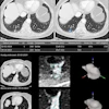





![Examples of ultrasound findings and techniques. (A) Images in a 39-year-old male patient with a mass in the left thigh. The mass is heterogeneous on the B-mode US image (compared with the patient in D) and showed increased microvascularity (superb microvascular imaging [SMI]) and shear-wave elastography (SWE) values. Undifferentiated pleomorphic sarcoma was diagnosed at biopsy (with pleomorphic rhabdomyosarcoma in surgical specimen). (B) Images in an 18-year-old male patient with a mass in the left leg. The mass is hypoechoic on the B-mode image, with no other findings suggestive of malignancy. The lesion is in contact with the cortex of the tibia, which is slightly irregular. CT revealed a doubtful anteromedial tibial erosion. The microvascular study demonstrated high vascularization, suggestive of malignancy. Periosteal Ewing sarcoma was diagnosed with both histologic and immunohistochemical confirmation. (C) Images in a 69-year-old female patient with a lump growing on the outside of the left leg. Multiple SWE examinations were performed (please note the high values obtained in the measurements, whereas the color map highlights the stiffness relative to adjacent tissues). SMI showed areas of increased vascularization to target for sampling. Undifferentiated spindle cell sarcoma was diagnosed at biopsy, with residual leiomyosarcoma in the surgical specimen after neoadjuvant therapy. (D) Images in a 56-year-old female patient with a mass in the right thigh. The mass is heterogeneous at both B-mode ultrasound (similar to patient A) and MRI (coronal T2-weighted spectral attenuated inversion recovery [SPAIR]; T1-weighted pre-contrast and postcontrast imaging), which even shows uptake after the administration of paramagnetic contrast material, which is traditionally suggestive of malignancy. Low values at SMI and elastography are suggestive of benignity. Spindle cell lipoma was diagnosed at biopsy, with atypical spindle cell lipomatous tumor in the surgical specimen.](https://img.auntminnieeurope.com/mindful/smg/workspaces/default/uploads/2026/08/images-radiol250278fig2.APCFLSvX6p.jpg?auto=format%2Ccompress&dpr=2&fit=crop&h=167&q=70&w=250)

