
An Egyptian expert in CT scans of mummies has called into question whether a mummified woman identified in a Polish museum last year was in fact pregnant, as claimed by authors of an article published in the Journal of Archaeological Science.
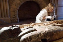
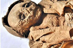
In early 2021, Polish researchers from the National Museum in Warsaw published a study based on their scans of what they said was a 25-year-old woman who lived in the Egyptian city of Thebes 3,000 years ago. At the time, they said the mummy was only known case of an embalmed pregnant woman.
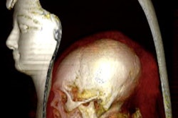
But those claims are being questioned by radiologist Dr. Sahar Saleem of Cairo University. She called for the authors of the study to rescan the mummy using proper protocol supervised by a paleoradiologist to clarify the diagnosis of the "pregnant mummy."
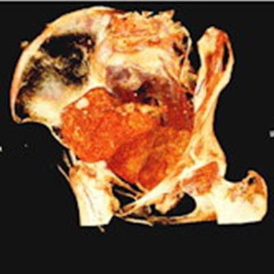
A second look could include possible differential diagnoses, such as visceral packs or condensed embalming materials, or a calcified pelvic tumor, Saleem suggested in her comments, published in the January issue of the Journal of Archaeological Science.
"Structures detected by CT in the pelvis of a mummy are usually embalming materials," Saleem wrote.
She noted that the scientific community and public should know that the discovery could be a pseudopregnancy.


![Overview of the study design. (A) The fully automated deep learning framework was developed to estimate body composition (BC) (defined as subcutaneous adipose tissue [SAT] in liters; visceral adipose tissue [VAT] in liters; skeletal muscle [SM] in liters; SM fat fraction [SMFF] as a percentage; and intramuscular adipose tissue [IMAT] in deciliters) from MRI. The fully automated framework comprised one model (model 1) to quantify different BC measures (SAT, VAT, SM, SMFF, and IMAT) as three-dimensional (3D) measures from whole-body MRI scans. The second model (model 2) was trained to identify standardized anatomic landmarks along the craniocaudal body axis (z coordinate field), which allowed for subdividing the whole-body measures into different subregions typically examined on clinical routine MRI scans (chest, abdomen, and pelvis). (B) BC was quantified from whole-body MRI in over 66,000 individuals from two large population-based cohort studies, the UK Biobank (UKB) (36,317 individuals) and the German National Cohort (NAKO) (30,291 individuals). Bar graphs show age distribution by sex and cohort. BMI = body mass index. (C) After the performance assessment of the fully automated framework, the change in BC measures, distributions, and profiles across age decades were investigated. Age-, sex-, and height-adjusted body composition reference curves were calculated and made publicly available in a web-based z-score calculator (https://circ-ml.github.io).](https://img.auntminnieeurope.com/mindful/smg/workspaces/default/uploads/2026/05/body-comp.XgAjTfPj1W.jpg?auto=format%2Ccompress&fit=crop&h=112&q=70&w=112)