Dear AuntMinnieEurope Member,
Being a Yorkshireman in the north of England, Dr. Chris Hammond doesn't go in for sugarcoating or superficiality. He gets straight to the point.
His latest column is a powerful, raw, and highly personal opinion piece about the memorable patients he's treated and the need to build resilience. It's a compelling read. Head over to the MRI Community for the full article.
The collapse of Christian Eriksen, Denmark's top-class midfielder, in last Saturday's Euros game against Finland was a chilling sight for anyone who saw it, according to renowned cardiac imaging expert Prof. Dr. Stephan Achenbach. He shared his thoughts with us about sudden cardiac death.
The UK Imaging & Oncology Congress has continued online this week, and we've got an informative and practical article from a leading children's hospital about how pediatric imaging has changed over the past 15 months.
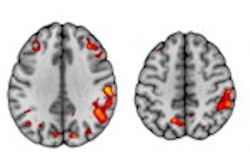
Meanwhile, Spanish researchers have shown that artificial intelligence-based analysis of 3D SPECT exams can help physicians to determine a patient's stage of Parkinson's disease. Their results deserve a close look in the Molecular Imaging Community.
Wherever possible, we try to post a short tribute to radiologists who've died of COVID-19. It's so important to ensure these people are remembered. The latest fatality is the 61-year-old celebrity radiologist Dr. Chinna Dua, who died in India on 11 June. Please contact me if you've lost any of your own radiology colleagues in the pandemic.


![Overview of the study design. (A) The fully automated deep learning framework was developed to estimate body composition (BC) (defined as subcutaneous adipose tissue [SAT] in liters; visceral adipose tissue [VAT] in liters; skeletal muscle [SM] in liters; SM fat fraction [SMFF] as a percentage; and intramuscular adipose tissue [IMAT] in deciliters) from MRI. The fully automated framework comprised one model (model 1) to quantify different BC measures (SAT, VAT, SM, SMFF, and IMAT) as three-dimensional (3D) measures from whole-body MRI scans. The second model (model 2) was trained to identify standardized anatomic landmarks along the craniocaudal body axis (z coordinate field), which allowed for subdividing the whole-body measures into different subregions typically examined on clinical routine MRI scans (chest, abdomen, and pelvis). (B) BC was quantified from whole-body MRI in over 66,000 individuals from two large population-based cohort studies, the UK Biobank (UKB) (36,317 individuals) and the German National Cohort (NAKO) (30,291 individuals). Bar graphs show age distribution by sex and cohort. BMI = body mass index. (C) After the performance assessment of the fully automated framework, the change in BC measures, distributions, and profiles across age decades were investigated. Age-, sex-, and height-adjusted body composition reference curves were calculated and made publicly available in a web-based z-score calculator (https://circ-ml.github.io).](https://img.auntminnieeurope.com/mindful/smg/workspaces/default/uploads/2026/05/body-comp.XgAjTfPj1W.jpg?auto=format%2Ccompress&fit=crop&h=112&q=70&w=112)


