
3D virtual reality (VR) simulation software can be a valuable pedagogical tool for radiography students, improving their confidence in performing a variety of radiographer tasks, according to research published online on 13 August in Radiography.
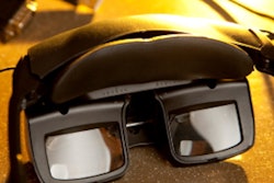
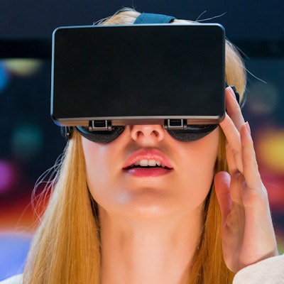
A team of researchers from University College Dublin in Ireland utilized commercial VR simulation technology to guide over 100 first-year radiography students through a comprehensive process of learning anatomy, radiographic positioning, and pathology. Next, the students x-rayed a virtual patient in the VR suite utilizing HTC Vive Pro headsets and hand controllers.
Of the 83 students who responded to an online survey afterward, 94% indicated they would recommend the VR software (Virtual Medical Coaching) to other students. Most (58%) said that they enjoyed the VR simulation, while 27% were indifferent. In addition, 58% wanted to have more access to VR, according to the authors led by Michelle O'Connor, PhD.
In terms of specific benefits, the students reported that VR practice improved their confidence in a number of areas:
- Beam collimation (75% of students)
- Centering of the x-ray tube (64%)
- Anatomical marker placement (63%)
- Exposure parameter selection (56%)
"3D immersive VR simulation is perceived by radiography students to be a valuable learning resource," they wrote. "VR needs to be strategically implemented into curricula to maximize its benefits."


![Overview of the study design. (A) The fully automated deep learning framework was developed to estimate body composition (BC) (defined as subcutaneous adipose tissue [SAT] in liters; visceral adipose tissue [VAT] in liters; skeletal muscle [SM] in liters; SM fat fraction [SMFF] as a percentage; and intramuscular adipose tissue [IMAT] in deciliters) from MRI. The fully automated framework comprised one model (model 1) to quantify different BC measures (SAT, VAT, SM, SMFF, and IMAT) as three-dimensional (3D) measures from whole-body MRI scans. The second model (model 2) was trained to identify standardized anatomic landmarks along the craniocaudal body axis (z coordinate field), which allowed for subdividing the whole-body measures into different subregions typically examined on clinical routine MRI scans (chest, abdomen, and pelvis). (B) BC was quantified from whole-body MRI in over 66,000 individuals from two large population-based cohort studies, the UK Biobank (UKB) (36,317 individuals) and the German National Cohort (NAKO) (30,291 individuals). Bar graphs show age distribution by sex and cohort. BMI = body mass index. (C) After the performance assessment of the fully automated framework, the change in BC measures, distributions, and profiles across age decades were investigated. Age-, sex-, and height-adjusted body composition reference curves were calculated and made publicly available in a web-based z-score calculator (https://circ-ml.github.io).](https://img.auntminnieeurope.com/mindful/smg/workspaces/default/uploads/2026/05/body-comp.XgAjTfPj1W.jpg?auto=format%2Ccompress&fit=crop&h=112&q=70&w=112)


