Chest CT examinations can play a critical role in the initial diagnosis and timely treatment of pneumonia caused by the novel coronavirus (2019-nCoV), according to an editorial published online on 6 February in European Radiology.
Recent reports have estimated the number of confirmed coronavirus cases to be more than 40,000 worldwide, with well over 900 deaths.
As 2019-nCoV can spread from person to person even among asymptomatic individuals, early diagnosis of the infection is crucial, wrote co-authors Yueying Pan and Hanxiong Guan from the radiology department at Huazhong University of Science and Technology in Wuhan, China. Chest CT plays a "very important" role in the initial diagnosis of coronavirus-infected pneumonia.
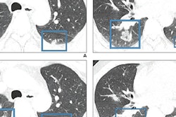
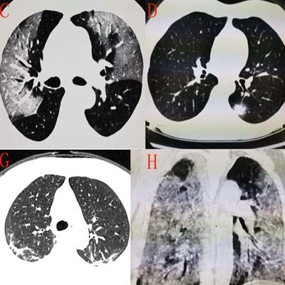
 CT scans of a patient with the novel coronavirus showing ground-glass opacities (A-C), pulmonary consolidation and nodules (D), local consolidation with peripheral ground-glass opacity (E), ground-glass opacities resembling "melted sugar" (F), inflammation absorbed by the lungs (G), and pneumonia in the white-lung stage (H). Figure courtesy of European Radiology.
"CT is often found to be positive when patients with 2019-nCoV develop persistent fever, cough, and unexplained weakness," they wrote.
In their editorial, the authors identified the most common CT features in coronavirus patients admitted to their hospital as well as the manner in which the features changed over time. They found that early CT lung manifestations were most often characterized by ground-glass opacities, pulmonary consolidation and nodules, and centralized local consolidation with ground-glass density. They also noted that, over time, lung consolidation tends to form into ground-glass opacities that appear like "melted sugar."
Treating patients at this stage of the disease generally leads to improvement, they wrote. Failing to treat the disease at this stage results in life-threatening consequences. The few patients examined in this latter stage of coronavirus-infected pneumonia had a "white lung" appearance on their CT scans.
"Early imaging diagnosis is of paramount importance," the authors concluded. "The purpose of this study is to help early diagnosis based on imaging findings, to set up early isolation and early treatment of patients, and to participate in controlling the outbreak by public health strategies of timely implementation of effective countermeasures."