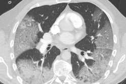
Although the imaging features of the novel coronavirus disease (COVID-19) overlap with those of severe acute respiratory syndrome (SARS) and Middle East respiratory syndrome (MERS), there are differences on imaging exams that set COVID-19 apart -- particularly that it tends to manifest in both lungs rather than one, according to a 28 February study in the American Journal of Roentgenology.
The new findings on how COVID-19 presents on imaging exams should help clinicians better diagnose the disease, wrote a team led by Dr. Melina Hosseiny of the University of California, Los Angeles (UCLA).
"Although the imaging features [of COVID-19] closely resemble those of MERS and SARS, involvement of both lungs on initial imaging is more likely to be seen with COVID-19," the group wrote. "Initial chest imaging abnormalities in SARS and MERS are more frequently unilateral."
SARS and MERS are also caused by coronaviruses. SARS was identified in 2003, and 8,422 patients were infected, with 916 deaths. It has been contained, the authors noted. MERS was first identified in 2012 and has been found in 2,492 patients, killing 858. New cases of MERS continue to arise, the latest being in December 2019.
Since the outbreak of COVID-19 in December of last year, more than 79,000 people have been infected around the world. As of February 24, more than 2,600 deaths from the virus have been recorded, according to Hosseiny and colleagues.
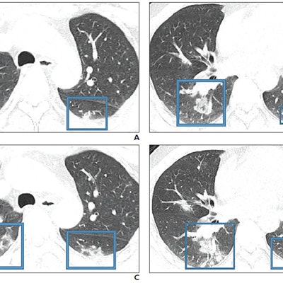
 47-year-old Chinese man with two-day history of fever, chills, productive cough, sneezing, and fatigue who presented to the emergency department. A and B: Initial CT images obtained show small round areas of mixed ground-glass opacity and consolidation (rectangles) at level of aortic arch (A) and ventricles (B) in right and left lower lobe posterior zones. C and D: Follow-up CT images obtained two days later show progression of abnormalities (rectangles) at the level of aortic arch (C) and ventricles (D), which now involve right upper and right and left lower lobe posterior zones. Images courtesy of the American Journal of Roentgenology.
The three diseases share the following symptoms: fever, dyspnea, malaise, myalgia, and headache; all three also tend to show ground-glass opacity, consolidation, or both on imaging, while lymphadenopathy is not usually seen. Clinicians have been diagnosing COVID-19 in patients with these symptoms who recently traveled to China.
"An investigation of initial chest CT findings in 21 individuals with confirmed COVID-19 reported abnormal findings in 86% of patients, with a majority having bilateral lung involvement," the authors wrote.
In addition to the bilateral lung involvement factor, another way the three viruses differ is in the features that indicate poor prognosis, according to Hosseiny's group:
- SARS: Bilateral (like acute respiratory distress syndrome), four or more lung zones, progressive involvement after 12 days
- MERS: Greater involvement of the lungs, pleural effusion, pneumothorax
- COVID-19: Consolidation (versus ground-glass opacity)
"Radiologists should be prepared for the incidence of COVID-19 to escalate," the investigators wrote. "Because the etiologic and clinical features of the syndrome are similar to those of SARS and MERS, the experience from those pulmonary syndromes can be helpful for managing the emerging COVID-19 outbreak."
Hosseiny and colleagues recommend patients recovering from COVID-19 be tracked with CT to assess for long-term or permanent lung damage, including fibrosis, as is seen in MERS and SARS infections.
"The experiences with SARS and MERS show that follow-up imaging should be performed in individuals recovering from COVID-19 to look for evidence of chronic involvement of the lungs (i.e., interlobular thickening, air trapping, or fibrosis)," the authors concluded.