The annual meeting of the European Society of Thoracic Imaging (ESTI), scheduled to take place in Istanbul from 26 to 28 May, has been called off due to security worries in the Turkish city. This setback follows the decision of the International Society of Radiology (ISR) to switch its 2014 congress from Cairo to Dubai, United Arab Emirates.
Soon after last week's bomb attack in which 10 German tourists were killed and another 17 people wounded in the historic center of Istanbul near the Blue Mosque, the society's president, Dr. Nevzat Karabulut, made the announcement of the cancellation in a message posted on the ESTI homepage. It was addressed to "colleagues, friends, and industry partners."
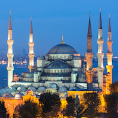
 The Blue Mosque (called Sultanahmet Camii in Turkish) was built between 1609 and 1616 and is a popular tourist destination in Istanbul.
"Based on the recent very unfortunate incident in Istanbul, Turkey, the executive board of ESTI and myself had to reconsider our annual scientific meeting 2016," he noted. "This decision was in no way easy and has been taken with deep regrets. The safety and security of all congress delegates, attendees, invited persons, and industry partners is of utmost importance, which is why we look forward to your understanding."
Karabulut is professor of radiology at Pamukkale University in Denizli, Turkey. He took his fellowship in chest radiology at the Medical College of Wisconsin in Milwaukee, U.S., between 1997 and 1998, and in 2001-2002 he did a research fellowship in abdominal MRI at the Medical West Virginia University's department of radiology.
“It is rather sad to witness how external conditions influence our scientific activities. Istanbul has been one of the most attractive meeting locations in the ESTI community until the recent unfortunate incident," he told AuntMinnieEurope.com in an email. "Even more bothersome is that the whole of Europe today is less safe than in the past as the terrorism is getting global."
"We must stand together and show our solidarity to maintain and continue our educational and scientific activities. In this regard, we are searching for the possibilities to hold an ESTI meeting and/or workshop in autumn in a safer place. We will announce it as soon as we decide the location and venue," he added.
Typically, ESTI members spend at least 50% of their time working in thoracic radiology and have published fairly extensively in this field. The society was founded in 1993, following a small group of European chest radiologists' meeting in Grenoble, France, in 1992.
 "We must stand together and show our solidarity," Dr. Nevzat Karabulut said.
The schedule for ESTI 2016 was to start the rating of abstracts at the beginning of February. Abstract classification was planned for mid-February, followed by notification of accepted abstracts at the end of the month. Poster submissions were also due to begin at the end of February, and a precongress workshop was scheduled for 25 May.
"Success in previous years proves the importance of, and interest in, thoracic imaging and the society is very much looking forward for welcoming everybody in the near future again to our important and successful scientific events," Karabulut wrote, adding that any questions should be sent to the ESTI office in Vienna ([email protected], Tel. +43 1 5322165).
The main activities of ESTI have focused on the annual meeting held in the country of the society's president. According to the ESTI website, the scientific programs of such meetings have included most advances in imaging techniques and scientific knowledge for chest diseases, and CME programs have been delivered by "the best European faculty in thoracic imaging." Beyond the annual meeting, the aims of ESTI include to provide recommendations and guidelines for appropriate practice in chest imaging, and to establish long-term relationships with industry partners in order to convert technologic innovations into new clinical applications.