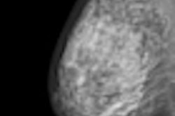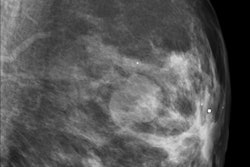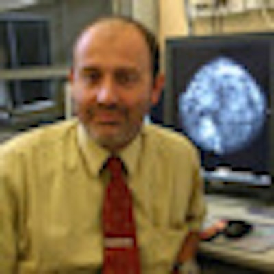
Hungary was a relatively early adopter when it came to launching a nationwide breast screening program, but this shouldn't come as a massive shock because the country holds some scientific ownership of the process. Dr. László Tabár, who led the landmark trials in Sweden 30 years ago, on which virtually all national screening programs are based, was born, educated, and trained in Hungary.
Based on a pilot scheme launched in 1995 that was partly funded by the World Bank, national screening was introduced to Hungary in 2002; neighboring Austria, by comparison, still doesn't have a countrywide program. The Hungarian program targets women between ages 45 and 65 and looks to screen patients biennially.
"Women are invited to attend by a personal letter, with appearance rates, depending on how you do the calculation, at 50% to 60%," explained Gábor Forrai, PhD, head of the radiology department at the ÁEK military hospital in Budapest. "It's not as high as it should be. We'd like it to be at least around 70%."
 Hungary also offers a dedicated training examination to radiolgoists innvolved in breast screening. All images courtesy of Gábor Forrai, PhD.
A radiographer conducts the clinical examination, which involves breast palpation and the taking of four images, two of each breast. "These are double read by two independent radiologists. If a suspicion of cancer is formed, the patient is called back and we will look at magnified views, perform ultrasound, and, if needed, biopsy," he said.
Forrai estimates that based on the screening results, perhaps 5% of women will be called back for further checks, usually at least for an ultrasound scan. "The figures are fairly standardized throughout the industrialized Western world; between one in eight and one in 10 women will develop breast cancer. In Hungary the figure is one in nine."
Given the Hungarian scheme has only been running 10 years, these are still early days. "Statistically, it is a very short period of time to be drawing firm conclusions, but the trends look good. What we observe from the latest stats is that breast cancer mortality rates are falling, mainly, I believe, because of our work," he said. In 2001, the number of breast cancer deaths among women in Hungary was 2,342. By 2007, the latest year for which detailed figures are available from Hungary's Central Statistical Office, it had fallen to 2,066.
"We are happy with the system, professionally speaking. Of course, you can always improve. Officials should be more flexible in fine-tuning; there are some small things that we still haven't been able to change after 10 years. But it is one of the success stories of Hungarian healthcare, and there aren't too many others," commented Forrai, a board member of the European Society of Breast Imaging (EUSOBI) who earlier this month spoke at a three-day course by the European Society of Radiology (ESR). "And it is a recognized scheme; we have a good reputation among the European radiology profession, too."
 EUSOBI plans to develop its own diploma, based in part on the Hungarian, U.K., Irish, and French experiences, according to Forrai.
Hungary also offers a dedicated training examination to radiologists innvolved in breast screening. "It exists in very few countries -- the U.K., the Republic of Ireland, and France," he said. "I don't know of any others in Europe, and the U.S. certainly doesn't have this."
The idea is twofold: Give radiologists the assurance that they really do know their science, and maintain public confidence in the system. That's important because the higher the patient turnout, the more effective the program will be. And breasts are notoriously difficult organs to read, according to Forrai. Get it wrong and either a cancer is missed or someone undergoes unnecessary surgery.
The training is organized by the University of Szeged hospital in the south of the country. A multiple-choice exam comes first, followed by reading a film on a viewing box (using genuine cases), then ultrasound tests. Candidates are questioned about biopsies and how to handle the ultrasound machine. Finally, there is an oral examination with Dr. Zoltán Pentek and Forrai, in which a real, complicated case is discussed. Here the radiologists need to show their ability to manage a patient, and teamwork is vital. Candidates must demonstrate they know what options are available for surgery or therapy, and how the team would be managed to deal with a patient.
Forrai estimates 1,200 radiologists are registered with the Society of Hungarian Radiologists, with about 800 active radiologists. About 120 radiologists are involved in mammography. The special examination was launched in 2008, since then 80 radiologists -- including Pentek and Forrai -- have passed.
At the moment, the exam is purely voluntary, though the national society of radiology would like it to become compulsory by law. EUSOBI also plans to develop its own diploma, based in part on the Hungarian, U.K., Irish, and French experiences, though unlike the Hungarian scheme, it will probably not include practical testing, just written and oral examinations.