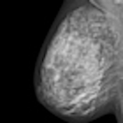
Fresh doubts have surfaced about the future of breast screening in the U.K. after a leading cancer specialist announced that an independent review of the National Health Service (NHS)'s program is under way.
"Should the independent review conclude that the balance of harms outweighs the benefits of breast screening, I will have no hesitation in referring the findings to the U.K. National Screening Committee and then (government) ministers," wrote Dr. Mike Richards, the national cancer director for Cancer and End of Life Care, Department of Health, London, in this week's British Medical Journal (BMJ 25 October 2011, online first). "I am fully committed to the public being given information in a format that they find acceptable and understandable and that enables them to make truly informed choices."
To resolve "the ongoing controversy" over breast screening, he has initiated a review of the research evidence that will be led by himself and Harpal Kumar, chief executive of Cancer Research U.K. Input will come from independent advisers who have never published on breast cancer screening. The evidence will then be presented at a workshop hosted by Cancer Research U.K., to which experts from both sides of the argument will be invited.
Mobile units are an integral component of the U.K. breast screening program, which is currently subject to an independent review.Richards also wants to establish a new process for developing written information for the public about each screening program, and plans to revise the current breast screening leaflet.
"As with any medical intervention, screening has potential risks that must be carefully evaluated against the benefits," he stated. "Work undertaken by eight leading international scientists calculated that the benefit of mammographic screening in terms of lives saved is greater than the harm in terms of overdiagnosis -- concluding that an estimated 2 to 2.5 lives are saved for every overdiagnosed case of breast cancer. I am, however, well aware that a contrary view has been provided by the Cochrane (Nordic Cochrane Centre) collaboration."
This announcement follows an open letter, also published by the BMJ (25 October 2011, online first), from Dr. Susan Bewley, professor of complex obstetrics in the division of women's health at King's College London. She thinks the harms of breast screening may outweigh the benefits and that screening information should be more balanced, and urged Richards to initiate a review of the evidence in light of growing uncertainty.
NHS leaflets have exaggerated the benefits of breast screening and do not spell out the risks, she noted. Furthermore, there is a growing body of international opinion acknowledging that breast cancer screening was not as good as used to be thought.
"The distress of overdiagnosis and decision-making when finding lesions that might (or might not) be cancer that might (or might not) require mutilating surgery is increasingly being exposed. The oft repeated statement that 1,400 lives a year are saved has not been subjected to proper scrutiny. Even cancer charities use lower estimates," she pointed out.
"There is a science behind the numbers and a duty to debate the facts, question the assumptions, and agree -- or at least explain systematically -- where the areas of dispute and uncertainty lie. How information is delivered must be explicit so that the ethical imperative for women to come to a free and fully informed decision is supported. The usefulness of screening diminishes with development of better treatments," Bewley added.
In his reply, Richards wrote screening programs should be based on the best available evidence, and based on current advice, including that of the World Health Organization, which says breast screening saves lives and the benefits considerably outweigh the harms. He also sought to reassure Bewley that he takes the current controversy very seriously.


![Examples of ultrasound findings and techniques. (A) Images in a 39-year-old male patient with a mass in the left thigh. The mass is heterogeneous on the B-mode US image (compared with the patient in D) and showed increased microvascularity (superb microvascular imaging [SMI]) and shear-wave elastography (SWE) values. Undifferentiated pleomorphic sarcoma was diagnosed at biopsy (with pleomorphic rhabdomyosarcoma in surgical specimen). (B) Images in an 18-year-old male patient with a mass in the left leg. The mass is hypoechoic on the B-mode image, with no other findings suggestive of malignancy. The lesion is in contact with the cortex of the tibia, which is slightly irregular. CT revealed a doubtful anteromedial tibial erosion. The microvascular study demonstrated high vascularization, suggestive of malignancy. Periosteal Ewing sarcoma was diagnosed with both histologic and immunohistochemical confirmation. (C) Images in a 69-year-old female patient with a lump growing on the outside of the left leg. Multiple SWE examinations were performed (please note the high values obtained in the measurements, whereas the color map highlights the stiffness relative to adjacent tissues). SMI showed areas of increased vascularization to target for sampling. Undifferentiated spindle cell sarcoma was diagnosed at biopsy, with residual leiomyosarcoma in the surgical specimen after neoadjuvant therapy. (D) Images in a 56-year-old female patient with a mass in the right thigh. The mass is heterogeneous at both B-mode ultrasound (similar to patient A) and MRI (coronal T2-weighted spectral attenuated inversion recovery [SPAIR]; T1-weighted pre-contrast and postcontrast imaging), which even shows uptake after the administration of paramagnetic contrast material, which is traditionally suggestive of malignancy. Low values at SMI and elastography are suggestive of benignity. Spindle cell lipoma was diagnosed at biopsy, with atypical spindle cell lipomatous tumor in the surgical specimen.](https://img.auntminnieeurope.com/mindful/smg/workspaces/default/uploads/2026/08/images-radiol250278fig2.APCFLSvX6p.jpg?auto=format%2Ccompress&fit=crop&h=100&q=70&w=100)


![Examples of ultrasound findings and techniques. (A) Images in a 39-year-old male patient with a mass in the left thigh. The mass is heterogeneous on the B-mode US image (compared with the patient in D) and showed increased microvascularity (superb microvascular imaging [SMI]) and shear-wave elastography (SWE) values. Undifferentiated pleomorphic sarcoma was diagnosed at biopsy (with pleomorphic rhabdomyosarcoma in surgical specimen). (B) Images in an 18-year-old male patient with a mass in the left leg. The mass is hypoechoic on the B-mode image, with no other findings suggestive of malignancy. The lesion is in contact with the cortex of the tibia, which is slightly irregular. CT revealed a doubtful anteromedial tibial erosion. The microvascular study demonstrated high vascularization, suggestive of malignancy. Periosteal Ewing sarcoma was diagnosed with both histologic and immunohistochemical confirmation. (C) Images in a 69-year-old female patient with a lump growing on the outside of the left leg. Multiple SWE examinations were performed (please note the high values obtained in the measurements, whereas the color map highlights the stiffness relative to adjacent tissues). SMI showed areas of increased vascularization to target for sampling. Undifferentiated spindle cell sarcoma was diagnosed at biopsy, with residual leiomyosarcoma in the surgical specimen after neoadjuvant therapy. (D) Images in a 56-year-old female patient with a mass in the right thigh. The mass is heterogeneous at both B-mode ultrasound (similar to patient A) and MRI (coronal T2-weighted spectral attenuated inversion recovery [SPAIR]; T1-weighted pre-contrast and postcontrast imaging), which even shows uptake after the administration of paramagnetic contrast material, which is traditionally suggestive of malignancy. Low values at SMI and elastography are suggestive of benignity. Spindle cell lipoma was diagnosed at biopsy, with atypical spindle cell lipomatous tumor in the surgical specimen.](https://img.auntminnieeurope.com/mindful/smg/workspaces/default/uploads/2026/08/images-radiol250278fig2.APCFLSvX6p.jpg?auto=format%2Ccompress&fit=crop&h=112&q=70&w=112)