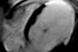
Using MRI, researchers have found that intra-aortic balloon pump counterpulsation prior to percutaneous coronary intervention in patients with ST segment elevation myocardial infarction does not reduce infarct size, according to a study presented on Tuesday at the European Society of Cardiology (ESC) congress in Paris.
Results from the Counterpulsation Reduces Infarct Size Acute Myocardial Infarction (CRISP AMI) trial found good results for intra-aortic balloon counterpulsation (IABC), according to principal investigator Dr. Manesh Patel from the Duke Clinical Research Institute of Duke University in Durham, NC.
IABC is a procedure in which a balloon is inserted in the aorta and is timed to inflate at the start of diastole and to deflate before the start of systole. The procedure increases diastolic pressure, which in turn increases coronary perfusion and delivers oxygen to the myocardium, facilitating the ejection of blood from the left ventricle.
Previous research has shown that left ventricular unloading before reperfusion can reduce the extent of the infarction. Infarct size has been known to predict left ventricular function after acute myocardial infarction.
Of the 337 patients enrolled in the multicenter prospective trial, 161 subjects randomly received IABC prior to primary percutaneous coronary intervention (PCI). The other 176 patients received standard care (primary PCI without IABC support).
Results showed that mean infarct size was not significantly different between the IABC and standard care groups (42% versus 37%, representing the percentage of left ventricle affected). At 30-day follow-up, major bleeding or transfusion had occurred in five (3%) IABC patients and three (2%) standard care patients.
Major vascular complications occurred in seven IABC (4%) and two standard care (1%) patients. At six months, death had occurred in three (2%) of the IABC and nine (5.2%) of the standard care group.
While the study did not meet its primary end point and was not powered to draw significant conclusions on clinical events, the "most striking observation is the excellent overall outcome for the highest risk patients within the context of this trial," Patel said. "The IABC group had less than 5% mortality, and it's difficult to improve that."
In the U.S., acute mortality risk in acute myocardial infarction is between 6% and 15%, with one-year mortality is estimated at 38% for women and 25% for men.


![Examples of ultrasound findings and techniques. (A) Images in a 39-year-old male patient with a mass in the left thigh. The mass is heterogeneous on the B-mode US image (compared with the patient in D) and showed increased microvascularity (superb microvascular imaging [SMI]) and shear-wave elastography (SWE) values. Undifferentiated pleomorphic sarcoma was diagnosed at biopsy (with pleomorphic rhabdomyosarcoma in surgical specimen). (B) Images in an 18-year-old male patient with a mass in the left leg. The mass is hypoechoic on the B-mode image, with no other findings suggestive of malignancy. The lesion is in contact with the cortex of the tibia, which is slightly irregular. CT revealed a doubtful anteromedial tibial erosion. The microvascular study demonstrated high vascularization, suggestive of malignancy. Periosteal Ewing sarcoma was diagnosed with both histologic and immunohistochemical confirmation. (C) Images in a 69-year-old female patient with a lump growing on the outside of the left leg. Multiple SWE examinations were performed (please note the high values obtained in the measurements, whereas the color map highlights the stiffness relative to adjacent tissues). SMI showed areas of increased vascularization to target for sampling. Undifferentiated spindle cell sarcoma was diagnosed at biopsy, with residual leiomyosarcoma in the surgical specimen after neoadjuvant therapy. (D) Images in a 56-year-old female patient with a mass in the right thigh. The mass is heterogeneous at both B-mode ultrasound (similar to patient A) and MRI (coronal T2-weighted spectral attenuated inversion recovery [SPAIR]; T1-weighted pre-contrast and postcontrast imaging), which even shows uptake after the administration of paramagnetic contrast material, which is traditionally suggestive of malignancy. Low values at SMI and elastography are suggestive of benignity. Spindle cell lipoma was diagnosed at biopsy, with atypical spindle cell lipomatous tumor in the surgical specimen.](https://img.auntminnieeurope.com/mindful/smg/workspaces/default/uploads/2026/08/images-radiol250278fig2.APCFLSvX6p.jpg?auto=format%2Ccompress&fit=crop&h=100&q=70&w=100)


![Examples of ultrasound findings and techniques. (A) Images in a 39-year-old male patient with a mass in the left thigh. The mass is heterogeneous on the B-mode US image (compared with the patient in D) and showed increased microvascularity (superb microvascular imaging [SMI]) and shear-wave elastography (SWE) values. Undifferentiated pleomorphic sarcoma was diagnosed at biopsy (with pleomorphic rhabdomyosarcoma in surgical specimen). (B) Images in an 18-year-old male patient with a mass in the left leg. The mass is hypoechoic on the B-mode image, with no other findings suggestive of malignancy. The lesion is in contact with the cortex of the tibia, which is slightly irregular. CT revealed a doubtful anteromedial tibial erosion. The microvascular study demonstrated high vascularization, suggestive of malignancy. Periosteal Ewing sarcoma was diagnosed with both histologic and immunohistochemical confirmation. (C) Images in a 69-year-old female patient with a lump growing on the outside of the left leg. Multiple SWE examinations were performed (please note the high values obtained in the measurements, whereas the color map highlights the stiffness relative to adjacent tissues). SMI showed areas of increased vascularization to target for sampling. Undifferentiated spindle cell sarcoma was diagnosed at biopsy, with residual leiomyosarcoma in the surgical specimen after neoadjuvant therapy. (D) Images in a 56-year-old female patient with a mass in the right thigh. The mass is heterogeneous at both B-mode ultrasound (similar to patient A) and MRI (coronal T2-weighted spectral attenuated inversion recovery [SPAIR]; T1-weighted pre-contrast and postcontrast imaging), which even shows uptake after the administration of paramagnetic contrast material, which is traditionally suggestive of malignancy. Low values at SMI and elastography are suggestive of benignity. Spindle cell lipoma was diagnosed at biopsy, with atypical spindle cell lipomatous tumor in the surgical specimen.](https://img.auntminnieeurope.com/mindful/smg/workspaces/default/uploads/2026/08/images-radiol250278fig2.APCFLSvX6p.jpg?auto=format%2Ccompress&fit=crop&h=112&q=70&w=112)