PARIS - Cardiac MRI is struggling to win a role in routine clinical use, but emerging research shows it holds powerful potential for advancing cardiologists' understanding of heart disease, noted speakers at the European Society of Cardiology (ESC) congress.
MRI is a complex technology -- more time-consuming, more expensive, and less available for everyday use than other modalities -- acknowledged Dr. Massimo Lombardi, from the Clinical Physiology Institute in Pisa, Italy. Yet his research on imaging of bicuspid aortic valves (BAV) attracted praise from the moderator of Tuesday's ESC "Imaging: Toy or tool?" session.
"Cardiovascular disease is so diffused, but it is clear that MRI and the molecular tools being developed are enriching an appreciation for the mechanisms underlying cardiovascular disease," said Dr. Valentin Fuster, PhD, director of Mount Sinai Heart in New York City.
 |
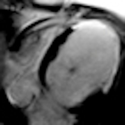
| MRI is leading to greater understanding of the mechanisms underlying cardiovascular disease. This figure shows postischemic dilated cardiomiopathy in a 58-year-old patient with known three-vessel coronary arteries disease. MR was planned to depict tissue viability prior to coronary revascularization. Left panel shows three chamber (upper row) and short axis (lower row) steady-state free precession cine MR images of an extremely dilated and remodeled left ventricle. On right panel, corresponding late-enhancement images show an extensive transmural area of enhancement involving the anterior, anteroseptal, and anterolateral midapical wall corresponding to extensive postischemic scar. MR findings suggest no chances of functional recovery after revascularization. Images courtesy of Dr. Marco Francone, University La Sapienza University, Rome. They originally appeared in ECR Today on 6 March 2011. |
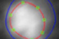
Lombardi's group has developed new techniques for measuring aortic wall biophysical properties for patients with BAV, a potentially lethal defect of the aortic valve that results in the formation of two leaflets or cusps instead of the normal three. BAV patients need to be regularly monitored, and the conventional examination using pulse wave velocity echocardiography effectively measures aortic compliance, the surrogate marker for determining changes that may indicate a deterioration of the condition.
"Echo is the first choice for its ability to precisely calculate this measure, but it does not give sufficient information and is operator-dependent, and results vary depending on the analysis software," he said.
In a paper published in July in the American Journal of Cardiology (1 July 2011, Vol. 108:1, pp. 81-87), Pisa researchers propose monitoring two new indexes for aortic compliance, the maximum rates of systolic distension and diastolic recoil (MRSD and MRDR). Fifty-three consecutive patients with BAV and healthy volunteers in a control group underwent a cardiac MR study that included phase velocity mapping and cine acquisition at several aortic levels.
Compared with the controls, MRSD was significantly lower in the whole BAV group where MRDR was greater. A curve analysis of MRSD distinguished BAV from controls with 100% sensitivity and 95% specificity, and both MRSD and MRDR were found to be slower in the patients with BAV than in the controls, regardless of the dimensions of the ascending aorta.
"We found a significant difference in these functions from the normal function of aortic valve and showed BAV is not just a disease of the valve but of the entire aortic function," Lombardi concluded.
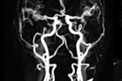
Much further upstream, researchers from the division of imaging sciences and biomedical engineering at King's College London are developing novel MR contrast agents and sequences for coronary vessel wall imaging.
Dr. Rene Botnar, chair of cardiovascular imaging, gave background to the paper, "Assessment of atherosclerotic plaque burden with an elastin-specific magnetic resonance contrast agent [ESMA]," published online in February in Nature Medicine (20 February 2011). Found in connective tissues, elastin content is especially high during plaque formation. Because ESMA binds to elastin and provides a strong signal for MRI, he was able to quantify intraplaque elastin content in a mouse model of atherosclerosis with high spatial resolution and accuracy. The study suggests a potential for developing a noninvasive assessment of plaque burden.
"This is the first time we were able to follow disease and this will probably lead to new insights into disease development," Botnar said, adding that techniques being developed for cardiac MRI are quite complicated and only 10% of them will make it to the clinic.