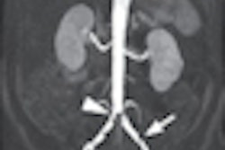
MONTREAL - With the use of a blood-pool contrast agent, MR angiography (MRA) can help simultaneously detect and assess arterial and additional venous conditions, according to a study presented on Monday at the International Society for Magnetic Resonance in Medicine (ISMRM) annual meeting.
Researchers from the University of Bonn found that incidental venous thrombosis was prevalent in 10% of patients who received MR angiography and the blood-pool contrast agent gadofosveset (Ablavar, Lantheus Medical Imaging) for suspected peripheral arterial occlusive disease.
The MRA-detected concomitant venous disease was not seen by standard first-pass imaging techniques. Lead study author and presenter Dr. Dariusch Hadizadeh said the ability to discover the additional maladies may have a much more positive effect on a patient's therapeutic management and treatment.
Longer imaging time
Currently, extracellular contrast agents are the primary diagnostic tool in many healthcare institutions for peripheral arterial occlusive disease. However, rapid diffusion of those agents reduces the vessel-to-tissue contrast over time, leaving a limited time window for image acquisition, Hadizadeh said.
By comparison, blood-pool contrast agents can provide image acquisition time of as much as one hour, offering high sensitivity and specificity for arterial disease evaluation and simultaneous visualization of arteries and veins.
The study examined 245 consecutive patients: 161 men, with a mean age of 64.3 years, and 84 women, with a mean age of 66.9 years. All patients were suspected of having arterial disease and underwent 1.5-tesla MRI (Achieva, Philips Healthcare) with gadofosveset.
For a control group, the researchers used contralateral veins with no evidence of disease, as examined by ultrasound (HD11, Philips) and the blood-pool contrast agent with MR angiography.
A flexible four-channel phased-array surface coil (SynBody, Philips) was used to image the lower legs. Images of the upper legs and the pelvic regions were acquired with the integrated quadrature body coil.
A total of 259 examinations were conducted, with 14 patients scanned twice during the study, resulting in a potential sample of 4,144 arterial segments. Forty-two segments were not assessable due to amputation of a leg, and one case of patient movement during the exam, leaving the researchers with 4,102 segments in the study sample.
In their analysis, Hadizadeh and colleagues found that most arterial segments (78%) had no relevant luminal stenosis (3,199 segments of stenosis less than 50%). There were 317 segments (8%) with occlusion of at least 50% and 586 arterial segments (14%) with total occlusion.
Surprising discovery
"We unexpectedly found 26 patients with incidental venous thrombosis," Hadizadeh said. "Acute thrombosis was found in 10 patients and 26 segments and chronic thrombosis was found in 17 patients and 35 segments."
Neither MR angiography with gadofosveset nor ultrasound detected thrombosis in any of the control veins, which included a total of 172 segments.
"The overall prevalence of incidental venous thrombosis in our peripheral arterial occlusive disease population was more than 10%," Hadizadeh said. "Acute thrombosis was found in 10 of 245 patients, and there was no significant relationship between the finding of deep venous thrombosis and peripheral arterial occlusive disease."


![Examples of ultrasound findings and techniques. (A) Images in a 39-year-old male patient with a mass in the left thigh. The mass is heterogeneous on the B-mode US image (compared with the patient in D) and showed increased microvascularity (superb microvascular imaging [SMI]) and shear-wave elastography (SWE) values. Undifferentiated pleomorphic sarcoma was diagnosed at biopsy (with pleomorphic rhabdomyosarcoma in surgical specimen). (B) Images in an 18-year-old male patient with a mass in the left leg. The mass is hypoechoic on the B-mode image, with no other findings suggestive of malignancy. The lesion is in contact with the cortex of the tibia, which is slightly irregular. CT revealed a doubtful anteromedial tibial erosion. The microvascular study demonstrated high vascularization, suggestive of malignancy. Periosteal Ewing sarcoma was diagnosed with both histologic and immunohistochemical confirmation. (C) Images in a 69-year-old female patient with a lump growing on the outside of the left leg. Multiple SWE examinations were performed (please note the high values obtained in the measurements, whereas the color map highlights the stiffness relative to adjacent tissues). SMI showed areas of increased vascularization to target for sampling. Undifferentiated spindle cell sarcoma was diagnosed at biopsy, with residual leiomyosarcoma in the surgical specimen after neoadjuvant therapy. (D) Images in a 56-year-old female patient with a mass in the right thigh. The mass is heterogeneous at both B-mode ultrasound (similar to patient A) and MRI (coronal T2-weighted spectral attenuated inversion recovery [SPAIR]; T1-weighted pre-contrast and postcontrast imaging), which even shows uptake after the administration of paramagnetic contrast material, which is traditionally suggestive of malignancy. Low values at SMI and elastography are suggestive of benignity. Spindle cell lipoma was diagnosed at biopsy, with atypical spindle cell lipomatous tumor in the surgical specimen.](https://img.auntminnieeurope.com/mindful/smg/workspaces/default/uploads/2026/08/images-radiol250278fig2.APCFLSvX6p.jpg?auto=format%2Ccompress&fit=crop&h=100&q=70&w=100)


![Examples of ultrasound findings and techniques. (A) Images in a 39-year-old male patient with a mass in the left thigh. The mass is heterogeneous on the B-mode US image (compared with the patient in D) and showed increased microvascularity (superb microvascular imaging [SMI]) and shear-wave elastography (SWE) values. Undifferentiated pleomorphic sarcoma was diagnosed at biopsy (with pleomorphic rhabdomyosarcoma in surgical specimen). (B) Images in an 18-year-old male patient with a mass in the left leg. The mass is hypoechoic on the B-mode image, with no other findings suggestive of malignancy. The lesion is in contact with the cortex of the tibia, which is slightly irregular. CT revealed a doubtful anteromedial tibial erosion. The microvascular study demonstrated high vascularization, suggestive of malignancy. Periosteal Ewing sarcoma was diagnosed with both histologic and immunohistochemical confirmation. (C) Images in a 69-year-old female patient with a lump growing on the outside of the left leg. Multiple SWE examinations were performed (please note the high values obtained in the measurements, whereas the color map highlights the stiffness relative to adjacent tissues). SMI showed areas of increased vascularization to target for sampling. Undifferentiated spindle cell sarcoma was diagnosed at biopsy, with residual leiomyosarcoma in the surgical specimen after neoadjuvant therapy. (D) Images in a 56-year-old female patient with a mass in the right thigh. The mass is heterogeneous at both B-mode ultrasound (similar to patient A) and MRI (coronal T2-weighted spectral attenuated inversion recovery [SPAIR]; T1-weighted pre-contrast and postcontrast imaging), which even shows uptake after the administration of paramagnetic contrast material, which is traditionally suggestive of malignancy. Low values at SMI and elastography are suggestive of benignity. Spindle cell lipoma was diagnosed at biopsy, with atypical spindle cell lipomatous tumor in the surgical specimen.](https://img.auntminnieeurope.com/mindful/smg/workspaces/default/uploads/2026/08/images-radiol250278fig2.APCFLSvX6p.jpg?auto=format%2Ccompress&fit=crop&h=112&q=70&w=112)