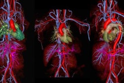
Time-resolved, or 4D, MR angiography (MRA) seems to be making rapid advances. The development of parallel-imaging techniques and hardware improvements such as gradient performance have enabled time-resolved MRA acquisition, which provides dynamic information at a feasible temporal resolution, along with a high spatial resolution, according to German researchers.
"Today 4D MRA techniques are increasingly applied in clinical settings," noted Nils Forkert, from the department of computational neuroscience at the University Medical Center Hamburg-Eppendorf in Hamburg, Germany. "New time-resolved MRA acquisition that does not require contrast agents has been proposed, and this makes it feasible to measure blood-flow velocity at different stages of the cardiac cycle."
In a presentation at the recent RSNA congress in Chicago, Forkert and his colleagues explained that an important drawback of classical 3D contrast-enhanced MRA has been the absence of temporal information. They reviewed three 4D MRA techniques according to their underlying imaging principles, their benefits and drawbacks, and their possible clinical applications for cerebrovascular diseases.
TWIST (4D time-resolved angiography with interleaved stochastic trajectories) depends on the fact that indicator substances cause a T1 shortening. The contrast agent is injected intravenously as a bolus, which dilutes downstream, where it is then measured using high temporal resolution MRI techniques. To achieve reasonable temporal resolutions, TWIST makes use of reduced echo and repetition times by asymmetric echo sampling and reduced k-space sampling using partial Fourier data sampling, rectangular fields-of-view, and parallel imaging. TWIST involves a one- to two-minute acquisition time, and full brain coverage is possible. Arterial and venous blood flow can be displayed, and neither ECG-synchronization nor special postprocessing tools are necessary. Conversely, multiple acquisitions are not possible, there is a trade-off between spatial and temporal resolution, and only indirect blood-flow velocity and direction information can be obtained, according to the authors.
Noncontrast-enhanced 4D MRA combines arterial spin labeling techniques with the STAR (signal targeting with alternating radiofrequency) flow-sensitive alternating inversion-recovery principle. The first dataset is acquired after upstream selective inversion, while the second is acquired without previous inversion. All spins that remain in the acquisition volume are saturated in both acquisitions and exhibit identical signals. Inflowing blood is only exposed to the upstream selective inversion pulse. Multiple acquisitions and full brain coverage are possible. No ECG synchronization is required, but it is recommended by Forkert, and postprocessing tools aren't necessary. A disadvantage is the long acquisition time of more than five minutes. Also, the image sequence is not broadly available yet, arterial and venous blood flow cannot be displayed at once, and only indirect blood flow velocity and information can be obtained.
Flow-sensitive 4D phase-contrast MRA aims to determine blood-flow velocities at different stages of the cardiac cycle. It applies oppositional aligned gradient pulses in x, y, and z directions. Resting spins receive no phase shift, while flowing spins exhibit a phase shift proportional to the velocity. No contrast agent is needed, and the technique can provide direct information about blood-flow velocity and direction, as well as full brain coverage and high spatial and temporal resolution. On the negative side, ECG-synchronization is required, the image sequence is not broadly available, arterial and venous blood flow cannot be displayed at once, the acquisition time is up to 10 minutes, and special postprocessing tools are necessary, the authors concluded.
|
Clinical applications of three leading 4D MRA techniques TWIST
Noncontrast-enhanced 4D MRA
Flow-sensitive 4D phase-contrast MRA
Source: Nils Forkert, University Medical Center Hamburg-Eppendorf, RSNA 2011 presentation. |