Specialization and superspecialization have made education and industrial activity more complex. As more products appear on the market, particularly those related to healthcare IT, interoperability must be tested. Furthermore, because an increasing variety of professionals are involved in patient care, modern teaching centers must embrace both medics and managers, basing their programs on the entire treatment process and a wide range of industrial products.
At the Academy for Medical Training and Simulation (AMTS) in Lucerne, Switzerland, medical and nonmedical healthcare professionals are able to learn and train in such a patient management process.
"AMTS is a neutral, interdisciplinary and interindustrial learning and meeting platform, capable of simulating diagnostic and therapeutic actions in a hospital-like environment," said surgeon and AMTS CEO Dr. Roger Zobrist.
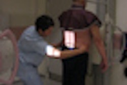
 Dr. Roger Zobrist (right) and colleague Dr. Ulrich Strycker prepare the simulator for emergency training. All images courtesy of AMTS.
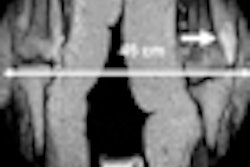
At the end of its first year of operation, AMTS has already seen about 4,500 course participants come through its doors, predominantly for musculoskeletal training, but also for events and courses in gastrointestinal and vascular surgery. Using real technical equipment and anatomic specimens, doctors can train without the need for live patients, which is particularly crucial for the imaging and surgical elements of the practical training.
Image guidance, including 3D visualization both pre- and intraoperatively, is increasingly important in all medical specialties. Such techniques are used throughout the medical process, from emergency admission to preoperative planning to intraoperative action. Knowledge is needed for the handling of image postprocessing software and documentation.
"Surgeons should be trained to use this software without the help of radiologists, despite the fact that in the era of image guidance, surgeons and radiologists should be partners and cooperate very closely," said Dr. Pietro Regazzoni, emeritus professor of surgery at the University of Basel, Switzerland, and a member of AMTS' board of directors.
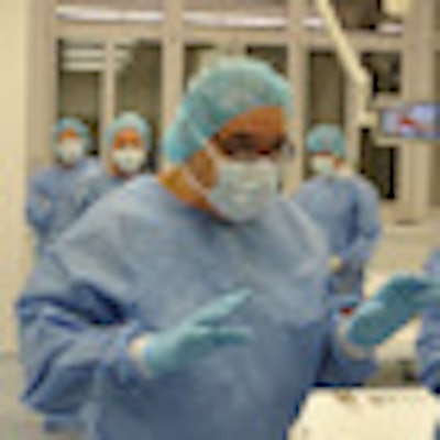
 A typical training session at AMTS. Dr. Pietro Regazzoni (shown in the center of the picture, with arms raised) believes industrial partners benefit from directly learning the needs of frontline healthcare staff and management through simulated work flow.
The contents of AMTS training will depend on future partners' needs and initiatives, as well those already in existence, such as the Swiss Telemedicine Society, which has its offices at the center. Telemedicine is a vast area for the center that includes image transfer, teleconsultation, second opinion, and monitoring of chronically ill patients. Its applications and corresponding products will be demonstrated and increasingly used at AMTS during the coming years, according to Regazzoni and Zobrist.
The learning platform of the academy also serves as an interindustrial meeting arena for its core business partners, they added. For information and communication technology, this function takes place through the Swiss e-Health Campus, an offspring company that plans to use AMTS' mocked-up hospital environment for interoperability testing. Medtech companies will be able to showcase and integrate their products at the academy, work on hybrid products needing input from more than one single company or industry, and provide in-house training for their own personnel and collaborators.
At the same time, medical and nonmedical decision-makers in the treatment process will get to see real solutions.
"Improvements in cost efficacy will depend on improvements to process quality. We want to encourage these new decision-makers to have their regular meetings here and to exchange views with other stakeholders," Regazzoni said.