Radiographers have an important role to play in monitoring patient radiation exposure and adjusting x-ray equipment settings when rates rise, according to Finnish researchers.
This was the lesson of a study using quality control measurements acquired by radiographers operating x-ray equipment at a health center in Haukipudas, a town of about 19,000 residents near the Gulf of Bothnia in northwestern Finland.
The researchers' findings identified sharply higher patient radiation exposure after the health service adopted a Finnish national policy recommending performing lumbar spine x-ray studies with the patient standing in a normal upright position. Radiation dosages later fell when a new scanner's automatic exposure control was tuned and radiographers placed more emphasis on manually adjusting its contrast and tube current settings to match patient body size.
Anja Henner, PhD, a principal lecturer at Oulu University of Applied Sciences in Oulu, Finland, presented the results at the 2011 ECR.
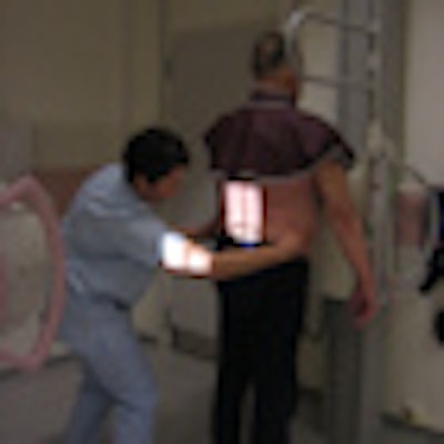
The radiographer's attention to imaging details, such as camber positioning, mAs, and kVp settings, have helped reduce radiation exposure during lumbar x-ray studies at the Haukipudas Health Center in northwest Finland. Image courtesy of Pöyskö Heli.
The framework for her study was set in the spring of 2009 when supervisors at Oulu University Hospital instructed two radiographers who perform x-ray procedures in its satellite health center in Haukipudas to adopt a different approach to lumbar x-ray imaging.
With the new policy, adult patients who complained of lumbar spine pain and were capable of standing without assistance were imaged on an upright wall-stand x-ray system. The previous policy called for examination of lumbar spine pain with the patient lying on a bucky table. Spine vertebra are more easily visualized while under the normal stress of supporting the patient in a standing position, Henner wrote in response to emailed questions from AuntMinnieEurope.
Radiation quality control measurements acquired annually from lumbar studies performed on at least 10 normal patients indicated the average dose area product for studies on the bucky table decreased steadily from 447.3 cGy cm2 in 2004 to 377.4 cGy cm2 in 2008 for anterior posterior and lateral views.
The downward trend ended abruptly in 2009 when the average jumped to 496.7 cGy cm2 after the center switched to the wall-stand camera.
But that peak was short-lived as staff addressed the problem, Henner noted. The average dose area product dropped to 350 cGy cm2 after the sensitivity of the system's automatic exposure control was increased to 80 kV for the anterior-posterior views and 90 kV for the lateral view.
Radiographers also focused more attention on patient size and weight when they manually adjusted mAs settings on the wall-stand system, she said. Additional refinements made after consultations with radiologists at the Oulu hospital helped drop the average dose area product to 208.4 cGy cm2.
Henner stressed that the entire process was possible because the radiographers were aware of dose levels at the clinic because they measured them regularly. Actions taken to reduce dosage were easy and inexpensive, she said.
"The key person in dose optimization is the radiographer," Henner said. "Working in this way demonstrates a very high commitment to the radiation safety culture."


![Examples of ultrasound findings and techniques. (A) Images in a 39-year-old male patient with a mass in the left thigh. The mass is heterogeneous on the B-mode US image (compared with the patient in D) and showed increased microvascularity (superb microvascular imaging [SMI]) and shear-wave elastography (SWE) values. Undifferentiated pleomorphic sarcoma was diagnosed at biopsy (with pleomorphic rhabdomyosarcoma in surgical specimen). (B) Images in an 18-year-old male patient with a mass in the left leg. The mass is hypoechoic on the B-mode image, with no other findings suggestive of malignancy. The lesion is in contact with the cortex of the tibia, which is slightly irregular. CT revealed a doubtful anteromedial tibial erosion. The microvascular study demonstrated high vascularization, suggestive of malignancy. Periosteal Ewing sarcoma was diagnosed with both histologic and immunohistochemical confirmation. (C) Images in a 69-year-old female patient with a lump growing on the outside of the left leg. Multiple SWE examinations were performed (please note the high values obtained in the measurements, whereas the color map highlights the stiffness relative to adjacent tissues). SMI showed areas of increased vascularization to target for sampling. Undifferentiated spindle cell sarcoma was diagnosed at biopsy, with residual leiomyosarcoma in the surgical specimen after neoadjuvant therapy. (D) Images in a 56-year-old female patient with a mass in the right thigh. The mass is heterogeneous at both B-mode ultrasound (similar to patient A) and MRI (coronal T2-weighted spectral attenuated inversion recovery [SPAIR]; T1-weighted pre-contrast and postcontrast imaging), which even shows uptake after the administration of paramagnetic contrast material, which is traditionally suggestive of malignancy. Low values at SMI and elastography are suggestive of benignity. Spindle cell lipoma was diagnosed at biopsy, with atypical spindle cell lipomatous tumor in the surgical specimen.](https://img.auntminnieeurope.com/mindful/smg/workspaces/default/uploads/2026/08/images-radiol250278fig2.APCFLSvX6p.jpg?auto=format%2Ccompress&fit=crop&h=100&q=70&w=100)


![Examples of ultrasound findings and techniques. (A) Images in a 39-year-old male patient with a mass in the left thigh. The mass is heterogeneous on the B-mode US image (compared with the patient in D) and showed increased microvascularity (superb microvascular imaging [SMI]) and shear-wave elastography (SWE) values. Undifferentiated pleomorphic sarcoma was diagnosed at biopsy (with pleomorphic rhabdomyosarcoma in surgical specimen). (B) Images in an 18-year-old male patient with a mass in the left leg. The mass is hypoechoic on the B-mode image, with no other findings suggestive of malignancy. The lesion is in contact with the cortex of the tibia, which is slightly irregular. CT revealed a doubtful anteromedial tibial erosion. The microvascular study demonstrated high vascularization, suggestive of malignancy. Periosteal Ewing sarcoma was diagnosed with both histologic and immunohistochemical confirmation. (C) Images in a 69-year-old female patient with a lump growing on the outside of the left leg. Multiple SWE examinations were performed (please note the high values obtained in the measurements, whereas the color map highlights the stiffness relative to adjacent tissues). SMI showed areas of increased vascularization to target for sampling. Undifferentiated spindle cell sarcoma was diagnosed at biopsy, with residual leiomyosarcoma in the surgical specimen after neoadjuvant therapy. (D) Images in a 56-year-old female patient with a mass in the right thigh. The mass is heterogeneous at both B-mode ultrasound (similar to patient A) and MRI (coronal T2-weighted spectral attenuated inversion recovery [SPAIR]; T1-weighted pre-contrast and postcontrast imaging), which even shows uptake after the administration of paramagnetic contrast material, which is traditionally suggestive of malignancy. Low values at SMI and elastography are suggestive of benignity. Spindle cell lipoma was diagnosed at biopsy, with atypical spindle cell lipomatous tumor in the surgical specimen.](https://img.auntminnieeurope.com/mindful/smg/workspaces/default/uploads/2026/08/images-radiol250278fig2.APCFLSvX6p.jpg?auto=format%2Ccompress&fit=crop&h=112&q=70&w=112)