Open MR examinations performed at specially equipped hospitals can help to overcome the hefty challenges posed by the growing number of excessively obese patients, according to German researchers.
MR machines are well-suited for imaging patients who exceed 150 kg because the radiofrequency can penetrate large amounts of adipose tissue. However, excessively obese patients cannot be examined in most conventional MR units due to the manufacturers' table weight restrictions or the conventional gantry diameter, which is too small for excessively obese bodies. New open systems have greater table weight capacity, and larger bore diameters or gaps bear the potential to examine and diagnose excessively obese patients.
"These extreme situations may only occur sporadically and should be performed in centers with the relevant expertise and experience," said Dr. Maximilian de Bucourt, a radiologist at the Charité Medical University in Berlin. "To create a more efficient and smoother workflow, an additional mobile MRI-compatible table would be useful in order to prepare an excessively obese patient outside the MR suite."
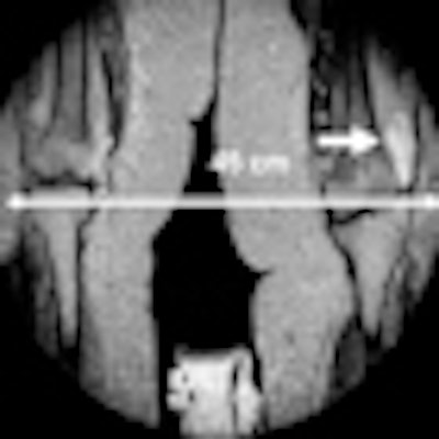
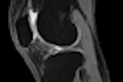
 T2-weighted, turbo spin-echo MR image with a field-of-view of 45 cm. The clinical findings of the excessively obese patient included pain in the left knee and laboratory values indicative of inflammation. MR depicted a hyperintense signal in the soft tissue lateral to the left femur condyle (white arrow). The soft-tissue abscess was confirmed by microbiology. Image courtesy of Dr. Maximilian de Bucourt.
Examining excessively obese patients can be difficult and expensive in a general clinical setting because of the special transportation requirements, anesthesia, artificial ventilation, or simply positioning the patient inside the MRI system with a sufficient amount of staff, de Bucourt noted. For instance, an MRI-compatible crane may be used to elevate part of the obese patient's weight to indirectly increase the overall weight capacity of the system's table. A general university hospital is best able to perform these exams, he added.
In the May edition of European Radiology, de Bucourt and his colleagues describe their experiences of evaluating 26 excessively obese patients in an open MRI system with a 250-kg table weight capacity, 40-cm gap, and a width of 160 cm. CT, conventional MR, and ultrasound exams were impossible or inconclusive in these patients, who had a body mass index greater than 35 and an average age of 46. All exams were performed on a 1-tesla Panorama high-field open system (Philips Healthcare) with a lightweight, vertical field, superconducting magnet, and a built-in solenoid technology transmit/receiver quadrature body coil (Eur Radiol, May 2011, Vol. 21:5, pp. 1004-1015).
A total of 24 out of 26 (92%) exams could be completed. In one emergency case (330 kg), only cranial MRI was possible due to the excessive weight exerted on the table. The possibility of table collapse and danger to the patient led to the decision to discontinue the MR study. The other exam was discontinued because the patient reported intense and increasing back pain while lying still.
As a result of this research, radiologists at Charité now receive referrals from other radiology departments and physicians when they deal with excessively obese patients, de Bucourt said.
Overall, he is concerned that excessive obesity can limit clinical and radiographic evaluation and may impede an accurate diagnosis. A complete physical exam is often impossible, and this places greater reliance on imaging. Ultrasound and plain x-ray have major limitations due to the decreased penetration of the large amounts of adipose tissue. Furthermore, the increased scattered radiation and beam-hardening secondary to the massive amounts of fat will often degrade CT images and limit the available information. Furthermore, radiation doses are much greater in these patients.


![A normal mammogram confirmed by three-year radiologic follow-up illustrates reader-marked regions of interest (ROIs) during (A) unaided (round 1) and (B) artificial intelligence (AI)–assisted (round 2) reading. Each colored dot represents an ROI for recall by a human reader. Readers could mark more than one ROI per case, represented by multiple dots of the same color. During AI-assisted reading, the AI system displayed three visible prompts: two with suspicion of malignancy scores of 35% (left mediolateral oblique [L MLO] and craniocaudal [L CC]) and one with a suspicion of malignancy score of 10% (right craniocaudal [R CC]), shown as polygonal overlays. Without AI, six of 10 readers (60%) marked a false-positive ROI. With AI assistance, this fell to two of 10 (20%). R MLO = right mediolateral oblique.](https://img.auntminnieeurope.com/mindful/smg/workspaces/default/uploads/2026/07/2026-07-14-radiology-mammogram-ai-auto-bias.H0bYO8QlWs.jpg?auto=format%2Ccompress&dpr=2&fit=crop&h=167&q=70&w=250)