

AuntMinnieEurope
  • Clinical News
  • Informatics
  • Industry News
  • Medical, Legal, And Practice
  • Education
  • Subspecialties
  • More
  • Sign In
  • Careers
  • Cases
  • Conferences
  • Videos
  • Webinars
  • Vendors
  • Advertising
  • AuntMinnie
Clinical News
  • CT
  • Digital X-Ray
  • Interventional
  • Molecular Imaging
  • MRI
  • Radiation Oncology/Therapy
  • Ultrasound
  • Womens Imaging
Imaging Informatics
  • Advanced Visualization
  • Enterprise Imaging
  • Artificial Intelligence
  • Cybersecurity
Industry News
  • Product News
  • Regulatory
  • M&A
  • Market Analysis
  • Installations
Medical, Legal, and Practice
  • Socioeconomic
  • Organized Radiology Issues
  • Service
Resources
  • Careers
  • Cases
  • Conferences
  • Videos
  • Webinars
  • Vendors
  • Advertising
  • AuntMinnie
User Tools
  • Sign In
More
  • About Us
  • Advertising
  • Help
  • AMEU Editorial Advisory Board
Follow AuntMinnieEurope
Facebook iconTwitter X icon LinkedIn iconInstagram icon
  • Clinical News
    • CT
    • Digital X-Ray
    • Interventional
    • Molecular Imaging
    • MRI
    • Radiation Oncology/Therapy
    • Ultrasound
    • Womens Imaging
  • Informatics
    • Advanced Visualization
    • Enterprise Imaging
    • Artificial Intelligence
    • Cybersecurity
  • Industry News
    • Product News
    • Regulatory
    • M&A
    • Market Analysis
    • Installations
  • Medical, Legal, And Practice
    • Socioeconomic
    • Organized Radiology Issues
    • Service
  • Education
    • Medical Students
    • Residents/Fellows
  • Subspecialties
    • Breast Imaging
    • CV
    • Chest
    • Emergency
    • GI
    • GU
    • Head & Neck
    • Interventional
    • Physics
    • MSK
    • Neuro
    • Nuclear
    • Pediatric
    • Radiation Oncology
  • More
  • Careers
  • Cases
  • Conferences
  • Videos
  • Webinars
  • Vendors
  • Advertising
  • AuntMinnie
  • Sign In
Follow AuntMinnieEurope
Facebook iconTwitter X icon LinkedIn iconInstagram icon

Top Story

Laim
Home
Leimgruber: Nuclear medicine needs regulation fit for innovation
In a video interview, Dr. Antoine Leimgruber, director ofdevelopment for nuclear medicine at Swiss Medical Network in Genolier, Switzerland, explores what is already changing in nuclear medicine and what continues to hold the specialty back.
Featured
Suhny
Home
Suhny Abbara talks AI in scientific publishing
Dr. Paul McCoubrie
Clinical News
The retiring radiologist
Latest News
Shutterstock 2437656775
SPECT, PET refine contemporary cardiac amyloidosis assessment
August 24, 2026
Examples of ultrasound findings and techniques. (A) Images in a 39-year-old male patient with a mass in the left thigh. The mass is heterogeneous on the B-mode US image (compared with the patient in D) and showed increased microvascularity (superb microvascular imaging [SMI]) and shear-wave elastography (SWE) values. Undifferentiated pleomorphic sarcoma was diagnosed at biopsy (with pleomorphic rhabdomyosarcoma in surgical specimen). (B) Images in an 18-year-old male patient with a mass in the left leg. The mass is hypoechoic on the B-mode image, with no other findings suggestive of malignancy. The lesion is in contact with the cortex of the tibia, which is slightly irregular. CT revealed a doubtful anteromedial tibial erosion. The microvascular study demonstrated high vascularization, suggestive of malignancy. Periosteal Ewing sarcoma was diagnosed with both histologic and immunohistochemical confirmation. (C) Images in a 69-year-old female patient with a lump growing on the outside of the left leg. Multiple SWE examinations were performed (please note the high values obtained in the measurements, whereas the color map highlights the stiffness relative to adjacent tissues). SMI showed areas of increased vascularization to target for sampling. Undifferentiated spindle cell sarcoma was diagnosed at biopsy, with residual leiomyosarcoma in the surgical specimen after neoadjuvant therapy. (D) Images in a 56-year-old female patient with a mass in the right thigh. The mass is heterogeneous at both B-mode ultrasound (similar to patient A) and MRI (coronal T2-weighted spectral attenuated inversion recovery [SPAIR]; T1-weighted pre-contrast and postcontrast imaging), which even shows uptake after the administration of paramagnetic contrast material, which is traditionally suggestive of malignancy. Low values at SMI and elastography are suggestive of benignity. Spindle cell lipoma was diagnosed at biopsy, with atypical spindle cell lipomatous tumor in the surgical specimen.
Advanced ultrasound techniques improve soft tissue tumor evaluation
August 26, 2026
Antoine Leimgruber
Swiss nuclear medicine seeks rules fit for radiopharmaceutical innovation
August 27, 2026
Radiotherapy 400
Radiotherapy access could address gynecological cancer disparities
August 24, 2026
Cases of the Week
Check out our Cases of the Week!
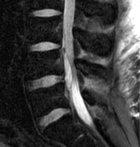
83-year-old female former smoker with imaging follow-up revisited
An 83-year-old woman with a history of prior tobacco use and pulmonary mycobacterial infection.
15-year-old boy with hemoptysis revisited
80-year-old man with altered mental status, seizure revisited
View All Cases
Clinical News
Shutterstock 2437656775
Nuclear Medicine
SPECT, PET refine contemporary cardiac amyloidosis assessment
Ultrasound
Advanced ultrasound techniques improve soft tissue tumor evaluation
Nuclear Medicine
Swiss nuclear medicine seeks rules fit for radiopharmaceutical innovation
Home
Leimgruber: Nuclear medicine needs regulation fit for innovation
Radiation Oncology/Therapy
Radiotherapy access could address gynecological cancer disparities
View More »
AI
Pexels Cottonbro 5867701 (1)
AI
Review highlights opportunities and risks of AI in pediatric imaging
Womens Imaging
AI performs well in breast screening, but density complicates the picture
AI
EU AI Act brings radiology AI oversight into daily practice
AI
Radiologists can identify three out of four AI-generated images
Womens Imaging
Incorrect AI suggestions influence reader performance on mammography
View More »
Industry News
Business Handshake 400
Industry News
Nanox AI signs distribution deal for HealthOST software
Industry News
Radiology Partners to acquire Everlight Radiology in $1 billion deal
Industry News
Philips, Imricor launch MRI-guided cardiac intervention lab platform
AI
Nanox AI signs UK distribution deal for HealthOST bone health software
Industry News
Ventripoint expands access to cardiac imaging platform
View More »
More from AuntMinnieEurope
Nanox AI signs distribution deal for HealthOST software
By AuntMinnie.com staff writers
The distribution deal with Vertec Scientific covers an initial three-year term.
August 27, 2026
Business Handshake 400
Radiology Partners to acquire Everlight Radiology in $1 billion deal
By AuntMinnie.com staff writers
Radiology Partners said it intends to make its Mosaic Drafting AI tool available to Everlight radiologists.
August 25, 2026
Acquisitions
Philips, Imricor launch MRI-guided cardiac intervention lab platform
By AuntMinnie.com staff writers
The platform combines Philips' 1.5T MRI system with Imricor's MR-compatible systems for MR-guided cardiac ablation.
August 27, 2026
Philips Rsna 2021 400
How much surveillance imaging is needed for meningiomas?
By Amerigo Allegretto
Most surveillance MRI or CT exams did not result in intervention, and treatment rates remained stable during the study period.
August 21, 2026
Mri Slide 400
Review charts path toward standardized chest x-ray optimization
By Claudia Tschabuschnig
A 29-study review found no consensus on optimal beam quality for adult chest radiography, prompting calls for standardized research and safer, more consistent protocols.
August 19, 2026
Shutterstock 2474798495
Fairness-aware training improves pneumonia AI performance across sexes
By Claudia Tschabuschnig
Fairness-aware training reduced several sex-based disparities in pneumonia AI detection without compromising overall accuracy, but widened the sensitivity gap.
August 19, 2026
Shutterstock 2743841919
AI framework could improve MRI quality control in prostate cancer
By Amerigo Allegretto
The framework uses a DL model trained on low- and high-quality MR images and helped improve radiologist accuracy.
August 19, 2026
Low-quality axial T2-weighted MRI scans with gradient-weighted class activation mapping. The low-quality MRI scans (top row) show blurred structures and artifacts making zonal and capsule boundaries indistinguishable. On the same scans with gradient-weighted class activation map overlays (bottom row), red indicates regions that were most heavily weighted by the deep learning model toward the low-quality prediction.
AI fracture detection device validated across three European hospitals
By Will Morton
No differences in AI performance were observed across centers, and AI assistance was associated with higher reader sensitivity.
August 18, 2026
Artificial intelligence (AI) assistance facilitates correct fracture identification in cases initially missed by human readers. A representative case is shown using anteroposterior radiographs in a 29-year-old male patient with wrist trauma (suspected nondisplaced transverse scaphoid fracture). The reader’s initial unaided assessment (top) resulted in a missed fracture, whereas with AI assistance (bottom), the fracture was correctly identified. The green box represents the fracture location as predicted by the AI tool, the blue box represents the reference standard annotation, and the red dots represent individual reader annotations.
Nanox AI signs UK distribution deal for HealthOST bone health software
By AuntMinnieEurope.com staff writers
HealthOST analyzes routine CT scans for spine assessment.
August 18, 2026
Nanox Logo 4
CT for breast radiotherapy detects ischemic heart disease risk
By Will Morton
As survival after early-stage breast cancer improves, cardiovascular disease has emerged as an increasingly prominent competing risk in older women.
August 17, 2026
Heart 1
Review highlights opportunities and risks of AI in pediatric imaging
By Claudia Tschabuschnig
Adult-trained AI tools may underperform in children, requiring pediatric-specific datasets, age-stratified validation, and stronger clinical oversight for safe deployment, researchers say.
August 17, 2026
Pexels Cottonbro 5867701 (1)
FAPI-PET/CT shows promise in hepatopancreatobiliary cancers
By Will Morton
FAPI-PET/CT could potentially improve noninvasive cancer staging, and, ultimately, clinical decision making.
August 13, 2026
Shown is a 69-year-old man with a cholangiocarcinoma in the distal common bile duct. F-18 FDG-PET/CT was negative where Ga-68 FAPI-46 PET/CT showed high focal FAPI uptake in the liver hilum and surrounding lymph nodes. a-d: F-18 FDG-PET/CT, e-h: Ga-68 FAPI-46 PET/CT. a, b, e, f: Maximum intensity projections, c, g: axial fusion images, d, h: non-contrast enhanced CT.
AuntMinnieEurope
Topics
  • Clinical News
  • Informatics
  • Industry News
  • Medical, Legal, And Practice
  • Education
  • Subspecialties
Resources
  • Careers
  • Cases
  • Conferences
  • Videos
  • Webinars
  • Vendors
  • Advertising
  • AuntMinnie
More
  • About Us
  • Advertising
  • Help
  • AMEU Editorial Advisory Board
Follow AuntMinnieEurope
Facebook iconTwitter X icon LinkedIn iconInstagram icon
  1. Privacy Policy
  2. Privacy Settings
  3. Terms & Conditions
  4. Contact Us
  5. Site Map
  6. Home
© 2026 Science and Medicine Group. All rights reserved.