A new review by European radiologists warns that artificial intelligence tools developed for adult imaging perform poorly and unsafely in children due to anatomical, physiological, and disease pattern differences, requiring pediatric-specific datasets, age-stratified validation, and continued radiologist oversight rather than autonomous reporting.
- Adult-trained AI models show significantly reduced performance in children, particularly those under 2 years old, with some systems achieving as low as 19% sensitivity for pediatric tuberculosis detection.
- Deep-learning reconstruction in CT imaging has reduced radiation dose by one-third to two-thirds while maintaining diagnostic quality in pediatric patients.
- Pediatric diseases are often rare with small, fragmented datasets across institutions, limiting AI development and creating weaker commercial incentives for pediatric-labeled products.
- Radiologists must remain responsible for reviewing AI output in pediatric imaging, with autonomous reporting avoided until stronger clinical evidence and external validation are available.
- Recommended solutions include pediatric-specific datasets, age-stratified evaluation, shared registries, federated learning, and collaboration among clinicians, researchers, regulators, and industry partners.
European radiologists should not assume that AI tools developed for adults will work safely in children, according to a new narrative review. The paper draws on a 2026 European Radiology scoping review and global multisociety guidance co-signed by the European Society of Paediatric Radiology (ESPR).
“Pediatric imaging must not be viewed simply as a delayed extension of adult radiology AI,” the authors wrote in the Indian Journal of Radiology and Imaging on August 13.
The review was led by Dr. Amit Gupta, MD, FRCR, of the All India Institute of Medical Sciences in New Delhi, India. The authors examined literature published from January 2005 through May 2026, emphasizing recent reviews, society statements, consensus guidance, and clinically relevant validation studies.
The work was narrative and did not include a formal systematic review or meta-analysis.
Small datasets and weak commercial incentives
Children differ from adults in anatomy, physiology, disease patterns, and imaging conditions. Normal appearances change substantially from infancy through adolescence, while motion, limited cooperation, smaller body size, sedation requirements, and the need to minimize radiation exposure all affect acquisition and interpretation.
Pediatric diseases may also be uncommon, leaving developers with small, fragmented datasets spread across institutions, age groups, and imaging protocols.
Lower pediatric imaging volumes also create weaker commercial incentives, contributing to fewer pediatric-labeled products and slower clinical translation than in adult radiology.
These constraints mean adult-trained algorithms may not generalize safely. A 2026 scoping review published in European Radiology found reduced performance when adult-derived imaging models were applied to children, particularly those under 2 years.
Gupta and colleagues also noted that one adult-trained computer-aided detection system achieved sensitivity as low as 19% for pediatric tuberculosis triage on chest radiographs.
Near-term opportunities
The authors identified image acquisition and quality optimization as particularly promising. Deep-learning reconstruction and denoising have reduced CT radiation dose by approximately one-third to two-thirds while preserving diagnostic quality, according to studies included in the review. In MRI, AI-assisted acceleration, motion correction, and improved signal-to-noise ratio could shorten examinations and potentially reduce sedation.
Automated bone-age assessment is the most mature interpretive application, with more than a decade of clinical use. However, performance can still vary at the extremes of age and in atypical populations, according to the authors.
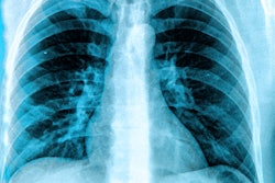
Pediatric chest radiograph interpretation is also widely studied, particularly for pneumonia, but external validation remains limited and many datasets mix adult and pediatric cases.
Address data scarcity
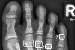
Other emerging or exploratory applications include fracture detection, abdominal imaging, neuroradiology, oncology radiomics, and chronic-lung-disease quantification. Their evidence bases are often retrospective, task-specific, and based on small cohorts.
The authors recommended pediatric-specific datasets, age-stratified evaluation, external and prospective validation, and postdeployment monitoring. Clinically meaningful outcomes should extend beyond diagnostic accuracy to radiation reduction, fewer sedations, successful examination completion, and equitable performance across age groups.
Shared registries, federated learning, standardized annotations, and collaboration among clinicians, researchers, regulators, industry, patients, and families could help address data scarcity. Until stronger evidence is available, radiologists should remain responsible for reviewing AI output, and autonomous reporting should be avoided.
The authors declared no conflicts of interest.
The full study can be found here.