Serial MR scanning shows that different components within carotid atherosclerotic plaque may influence each other and increase the risk of intraplaque hemorrhage (IPH), according to neurovascular imaging research published on 3 June in Radiology.
The authors were led by Dr. Daniel Bos, PhD, associate professor in epidemiology and radiology and principal investigator of imaging of arteriosclerosis at Erasmus MC University Medical Center in Rotterdam, the Netherlands. They found that compared with plaques without calcification, plaques that already had calcification were twice as likely to develop internal bleeding, a key indicator of plaque vulnerability and potential rupture. And men are more likely to have plaques evolve to multicomponent plaques with IPH, the team also found.
Knowledge about the evolution of carotid atherosclerotic plaque composition in its preclinical phase is becoming increasingly important, according to the group. A better understanding of plaque progression could facilitate earlier stroke prevention strategies.
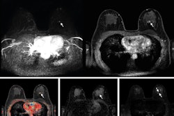
 Demonstration of colocalization of pre-existing calcification and incident intraplaque hemorrhage (IPH) in the same plaque. Axial cross sections of the same plaque at the same arterial location on a scan obtained with a T1-weighted gradient-echo noncontrast MRI sequence in a 70-year-old female participant who was free of coronary heart disease and stroke before follow-up MRI. Baseline (left) and follow-up (right) MRI examinations. The blue arrows highlight calcification in a hypointense signal area, while the green arrow indicates IPH as a hyperintense signal. The asterisks mark the lumen of the carotid artery.RSNA
The prospective study was embedded within the population-based Rotterdam Study, an ongoing population-based study recruiting residents aged 45 years and over in the Ommoord district of Rotterdam.
"Over the past decade, improvements in in vivo imaging techniques, especially MRI, have fueled the insight that atherosclerotic plaque components, as opposed to plaque size or luminal stenosis, are likely more important for determining the subsequent risk of ischemic stroke, particularly among persons with no or low-degree carotid stenosis," the group noted.
Beginning with 1,740 participants completing a first MRI examination between October 2007 and November 2012, Bos and colleagues followed 802 participants (1,460 arteries; mean age, 68.5 years; 461 male) who completed their follow-up scan six years later, between August 2014 and May 2017.
All participants were in the early stages of their disease, before any symptoms. Carotid MRI examinations were performed on the same 1.5-tesla MRI scanner (Signa Excite; GE HealthCare), following a standardized protocol, using a bilateral phased-array surface coil (Machnet, Eelde, the Netherlands).
The study measured carotid plaque compositions at baseline: 20.1% of plaques (294 of 1,460) were free of calcification, lipid-rich necrotic core, and IPH, whereas 28.4% (415 of 1,460) had two or more components. Carotid stenosis exceeding 50% was confirmed in 2.9% (43 of 1,460) at baseline and 3.3% (48 of 1,460) at follow-up.
Overall results showed that carotid plaques with calcification at baseline were independently associated with a higher incidence of IPH at follow-up (adjusted odds ratio, 2.00 [95% CI: 1.26, 3.16]; p = 0.003).
Compared with women, men were more likely to have plaques with no component or a single component evolve to multicomponent plaques with IPH (men, 21% [116 of 558]; women, 13% [61 of 468]; p < 0.001).
Additionally, simulating a 30-year evolution of plaque compositions, the group noted that multicomponent plaques represented 10% at age 55 years and increased to over 50% after age 70 years.
The authors acknowledged several limitations of the study, including that their noncontrast MRI examinations could not provide accurate information about the fibrous cap of carotid plaques, which is also an important plaque component. Also, the plaque components were assessed qualitatively without considering volume or morphologic characteristics.
Regardless, "one of the key findings of our work is that calcified plaques may not be as harmless as once thought, since these plaques were found to be at risk of intraplaque bleeding, which in itself is the most important cause of plaque rupture and subsequent stroke," Bos stated for the RSNA. He emphasized the importance of ongoing plaque monitoring and proactive risk factor management.
The group also called for future longitudinal research to confirm whether the morphologic characteristics of calcification are linked to incident IPH.
For more details on the study, go to press.rsna.org/timssnet/media/pressreleases/14_pr_target.cfm?ID=2583