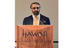
U.K.-trained head and neck radiologist Dr. Salman Qureshi has unveiled his social media channels about the importance of wellness, weight loss, lifestyle, and travel.
In a video interview with AuntMinnieEurope.com posted on 18 March, Qureshi spoke about his relocation from the U.K. to the Middle East and his personal health and fitness transformation that involved losing 43 kg (nearly 7 stone) in four years.
“Transformation is not just about appearance. It is about clarity, consistency, and long-term change,” he says on his LinkedIn page. “Approaching my 50s, I chose to prioritise my health. After years of working in brain imaging, I realised it was time to align my own lifestyle with what I knew from science.”
“They said it’s too late at 50. I disagreed...” notes Dr. Salman Qureshi.
The title of his YouTube channel is: Walking Changed My Physique & Opened A New Chapter | Fitness Transformation at 50. “It (the channel) is getting very good traction at the moment,” he pointed out on 19 May. “Even less than 24 hours in, all the videos combined have garnered between 3,000 and 4,000 views.”
Qureshi is a head and neck consultant and neuroradiologist at Sheikh Shakhbout Medical City (SSMC), Abu Dhabi, United Arab Emirates, and is a member of the Executive Committee of the European Society of Head & Neck Radiology. He previously worked at Hamad Medical Corporation in Doha, Qatar, and for seven years at Manchester University Foundation Trust.
Although he currently works in Abu Dhabi, he lives about 110 km (68 miles) away in Dubai, and many of his social media posts focus on Dubai.
Overall, his aim is to combine science and lifestyle – “revealing the extraordinary personal health transformation that sparked Doc Dubai’s passion to share knowledge and insight,” Qureshi notes on his website. “From brain-body connections to immersive travel content and the dynamic lifestyle of Dubai, Doc Dubai is here to inform, inspire, and entertain. Join us for a smarter, healthier, and more vibrant way of living.”
“They said it’s too late at 50. I disagreed…” he states in a YouTube episode.


![Overview of the study design. (A) The fully automated deep learning framework was developed to estimate body composition (BC) (defined as subcutaneous adipose tissue [SAT] in liters; visceral adipose tissue [VAT] in liters; skeletal muscle [SM] in liters; SM fat fraction [SMFF] as a percentage; and intramuscular adipose tissue [IMAT] in deciliters) from MRI. The fully automated framework comprised one model (model 1) to quantify different BC measures (SAT, VAT, SM, SMFF, and IMAT) as three-dimensional (3D) measures from whole-body MRI scans. The second model (model 2) was trained to identify standardized anatomic landmarks along the craniocaudal body axis (z coordinate field), which allowed for subdividing the whole-body measures into different subregions typically examined on clinical routine MRI scans (chest, abdomen, and pelvis). (B) BC was quantified from whole-body MRI in over 66,000 individuals from two large population-based cohort studies, the UK Biobank (UKB) (36,317 individuals) and the German National Cohort (NAKO) (30,291 individuals). Bar graphs show age distribution by sex and cohort. BMI = body mass index. (C) After the performance assessment of the fully automated framework, the change in BC measures, distributions, and profiles across age decades were investigated. Age-, sex-, and height-adjusted body composition reference curves were calculated and made publicly available in a web-based z-score calculator (https://circ-ml.github.io).](https://img.auntminnieeurope.com/mindful/smg/workspaces/default/uploads/2026/05/body-comp.XgAjTfPj1W.jpg?auto=format%2Ccompress&fit=crop&h=100&q=70&w=100)


![Overview of the study design. (A) The fully automated deep learning framework was developed to estimate body composition (BC) (defined as subcutaneous adipose tissue [SAT] in liters; visceral adipose tissue [VAT] in liters; skeletal muscle [SM] in liters; SM fat fraction [SMFF] as a percentage; and intramuscular adipose tissue [IMAT] in deciliters) from MRI. The fully automated framework comprised one model (model 1) to quantify different BC measures (SAT, VAT, SM, SMFF, and IMAT) as three-dimensional (3D) measures from whole-body MRI scans. The second model (model 2) was trained to identify standardized anatomic landmarks along the craniocaudal body axis (z coordinate field), which allowed for subdividing the whole-body measures into different subregions typically examined on clinical routine MRI scans (chest, abdomen, and pelvis). (B) BC was quantified from whole-body MRI in over 66,000 individuals from two large population-based cohort studies, the UK Biobank (UKB) (36,317 individuals) and the German National Cohort (NAKO) (30,291 individuals). Bar graphs show age distribution by sex and cohort. BMI = body mass index. (C) After the performance assessment of the fully automated framework, the change in BC measures, distributions, and profiles across age decades were investigated. Age-, sex-, and height-adjusted body composition reference curves were calculated and made publicly available in a web-based z-score calculator (https://circ-ml.github.io).](https://img.auntminnieeurope.com/mindful/smg/workspaces/default/uploads/2026/05/body-comp.XgAjTfPj1W.jpg?auto=format%2Ccompress&fit=crop&h=112&q=70&w=112)


