
Dear AuntMinnieEurope MRI Insider,
Automated quantification of tumor load to assess treatment response will become more important as systemic medical therapies evolve quickly. This is one of the key findings of new analysis by orthopedic surgeons and radiologists at Leiden University Medical Centre, in the Netherlands.
The researchers explain that emerging 3D MR segmentation techniques make it feasible to quantify tumor burden and provide a quick visual assessment of lesion distribution throughout the knee, but the process is time-consuming and operator-dependent. Their results deserve a close look in the MRI Community.
One of the ways multiple sclerosis (MS) manifests is as white-matter lesions in the brain, but the detection of new and enlarging white-matter lesions remains a challenging area in suspected cases of MS. In a five-center study involving MRI, Swiss investigators have evaluated the accuracy of software for finding these problematic lesions.
How can pelvic MRI be used to diagnose deep infiltrating endometriosis? How does the technique compare with transvaginal ultrasound? A research group from Pierre and Marie Curie University in Paris has addressed these questions in a recent publication.
When it comes to abdominal MRI, Prof. Luis Martí-Bonmatí from Valencia, Spain, is a recognized expert, so it's reassuring to know that he's closely involved in the European Federation for Cancer Images, a four-year project that aims to enhance the use of imaging and artificial intelligence technology for precision medicine in cancer patients.
In other news, Dr. Nathalie Hottat, a clinical radiologist at Burgmann University Hospital in Brussels, Belgium, and her colleagues have reported that diffusion-weighted MRI can help predict breast cancer patients' response to neoadjuvant chemotherapy.
The application of MR spectroscopy continues to be a promising area of advancement in brain tumor management, with significant benefits in the context of different physiological imaging techniques, according to researchers from Ireland.
In this letter, we've highlighted just a few of the many reports posted in the MRI Community over recent weeks. Please scroll through the full list below, and feel free to contact me if you have ideas for future coverage.

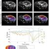
![Overview of the study design. (A) The fully automated deep learning framework was developed to estimate body composition (BC) (defined as subcutaneous adipose tissue [SAT] in liters; visceral adipose tissue [VAT] in liters; skeletal muscle [SM] in liters; SM fat fraction [SMFF] as a percentage; and intramuscular adipose tissue [IMAT] in deciliters) from MRI. The fully automated framework comprised one model (model 1) to quantify different BC measures (SAT, VAT, SM, SMFF, and IMAT) as three-dimensional (3D) measures from whole-body MRI scans. The second model (model 2) was trained to identify standardized anatomic landmarks along the craniocaudal body axis (z coordinate field), which allowed for subdividing the whole-body measures into different subregions typically examined on clinical routine MRI scans (chest, abdomen, and pelvis). (B) BC was quantified from whole-body MRI in over 66,000 individuals from two large population-based cohort studies, the UK Biobank (UKB) (36,317 individuals) and the German National Cohort (NAKO) (30,291 individuals). Bar graphs show age distribution by sex and cohort. BMI = body mass index. (C) After the performance assessment of the fully automated framework, the change in BC measures, distributions, and profiles across age decades were investigated. Age-, sex-, and height-adjusted body composition reference curves were calculated and made publicly available in a web-based z-score calculator (https://circ-ml.github.io).](https://img.auntminnieeurope.com/mindful/smg/workspaces/default/uploads/2026/05/body-comp.XgAjTfPj1W.jpg?auto=format%2Ccompress&fit=crop&h=100&q=70&w=100)


![Overview of the study design. (A) The fully automated deep learning framework was developed to estimate body composition (BC) (defined as subcutaneous adipose tissue [SAT] in liters; visceral adipose tissue [VAT] in liters; skeletal muscle [SM] in liters; SM fat fraction [SMFF] as a percentage; and intramuscular adipose tissue [IMAT] in deciliters) from MRI. The fully automated framework comprised one model (model 1) to quantify different BC measures (SAT, VAT, SM, SMFF, and IMAT) as three-dimensional (3D) measures from whole-body MRI scans. The second model (model 2) was trained to identify standardized anatomic landmarks along the craniocaudal body axis (z coordinate field), which allowed for subdividing the whole-body measures into different subregions typically examined on clinical routine MRI scans (chest, abdomen, and pelvis). (B) BC was quantified from whole-body MRI in over 66,000 individuals from two large population-based cohort studies, the UK Biobank (UKB) (36,317 individuals) and the German National Cohort (NAKO) (30,291 individuals). Bar graphs show age distribution by sex and cohort. BMI = body mass index. (C) After the performance assessment of the fully automated framework, the change in BC measures, distributions, and profiles across age decades were investigated. Age-, sex-, and height-adjusted body composition reference curves were calculated and made publicly available in a web-based z-score calculator (https://circ-ml.github.io).](https://img.auntminnieeurope.com/mindful/smg/workspaces/default/uploads/2026/05/body-comp.XgAjTfPj1W.jpg?auto=format%2Ccompress&dpr=2&fit=crop&h=167&q=70&w=250)


